3R1F image
Deposition Date 2011-03-10
Release Date 2011-08-03
Last Version Date 2024-10-16
Entry Detail
PDB ID:
3R1F
Keywords:
Title:
Crystal structure of a key regulator of virulence in Mycobacterium tuberculosis
Biological Source:
Source Organism:
Host Organism:
Method Details:
Experimental Method:
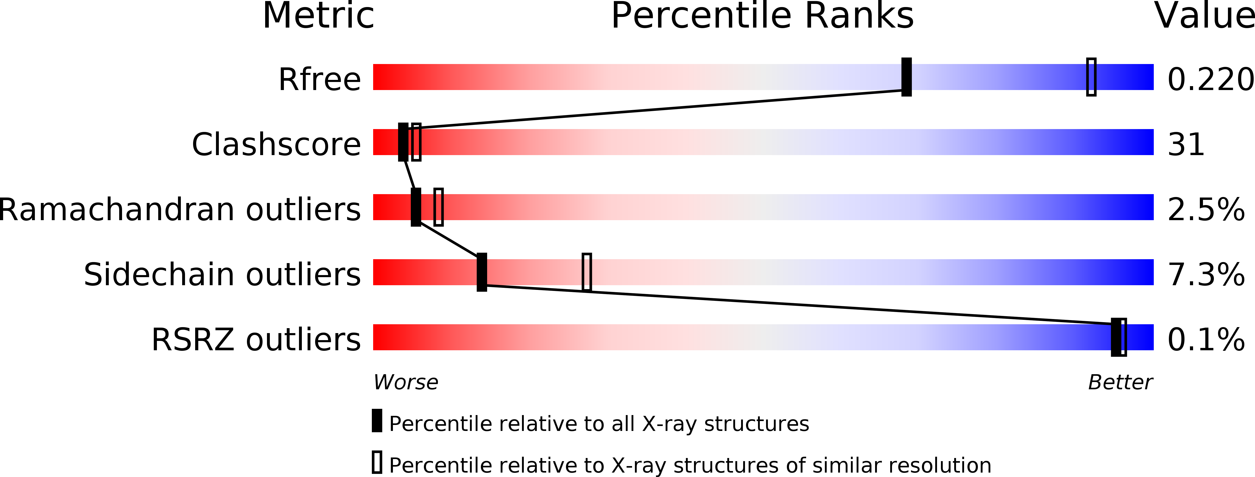
Resolution:
2.50 Å
R-Value Free:
0.23
R-Value Work:
0.19
R-Value Observed:
0.19
Space Group:
P 32
Macromolecular Entities
Polymer Type:polypeptide(L)
Molecule:ESX-1 secretion-associated regulator EspR
Chain IDs:A, B, C, D, E, F, G, H, I, J, K, L, M, N, O, P, Q, R
Chain Length:135
Number of Molecules:18
Biological Source:Mycobacterium tuberculosis
Modified Residue
Compound ID Chain ID Parent Comp ID Details 2D Image
MSE A MET SELENOMETHIONINE
Primary Citation
EspR, a key regulator of Mycobacterium tuberculosis virulence, adopts a unique dimeric structure among helix-turn-helix proteins.
Proc.Natl.Acad.Sci.USA 108 13450 13455 (2011)
PMID: 21795602 DOI: 10.1073/pnas.1110242108

Abstact

EspR is a transcriptional regulator that activates the ESX-1 secretion system during Mycobacterium tuberculosis infection and is critical for pathogenesis. It is unique among DNA-binding proteins as it is secreted as part of a feedback regulatory loop that serves to mitigate transcriptional activity. Here we report the crystal structure of a functional EspR dimer at 2.5-Å resolution. The amino-terminal half of EspR is a helix-turn-helix (HTH) DNA-binding domain and the carboxy terminus consists of a dimerization domain with similarity to the SinR:SinI sporulation regulator of Bacillus subtilis. Surprisingly, the HTH domains of EspR are arranged in an unusual conformation in which they are splayed at an oblique angle to each other, suggesting that EspR binds DNA in a profoundly different way than most other known HTH regulators. By mapping the EspR binding sites in the espACD promoter, using both in vivo and in vitro binding assays, we show that the EspR operators are located unusually far from the promoter. The EspR dimer binds to these sites cooperatively, but the two "half-sites" contacted by each DNA recognition motif are separated by 177 base pairs. The distinctive structure of EspR and the exceptional arrangement of its operator contacts suggest that it could promote DNA looping in its target promoter. We hypothesize that direct DNA looping mediated by single-site binding of each EspR monomer may facilitate transcriptional control of this important virulence system.

Legend

Protein

Chemical

Disease

Primary Citation of related structures