Deposition Date
2011-03-08
Release Date
2011-11-30
Last Version Date
2024-03-20
Entry Detail
PDB ID:
3R0H
Keywords:
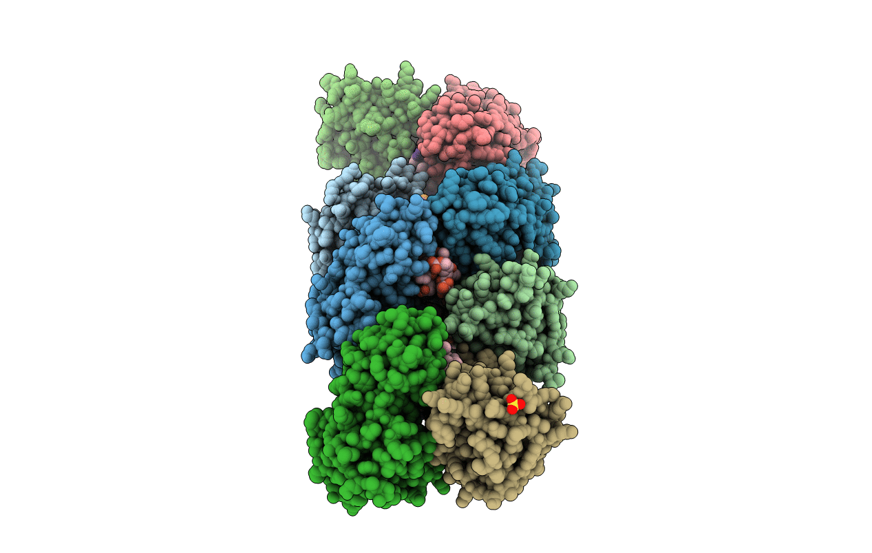
Title:
Structure of INAD PDZ45 in complex with NG2 peptide
Biological Source:
Source Organism(s):
Drosophila melanogaster (Taxon ID: 7227)
Expression System(s):
Method Details:
Experimental Method:
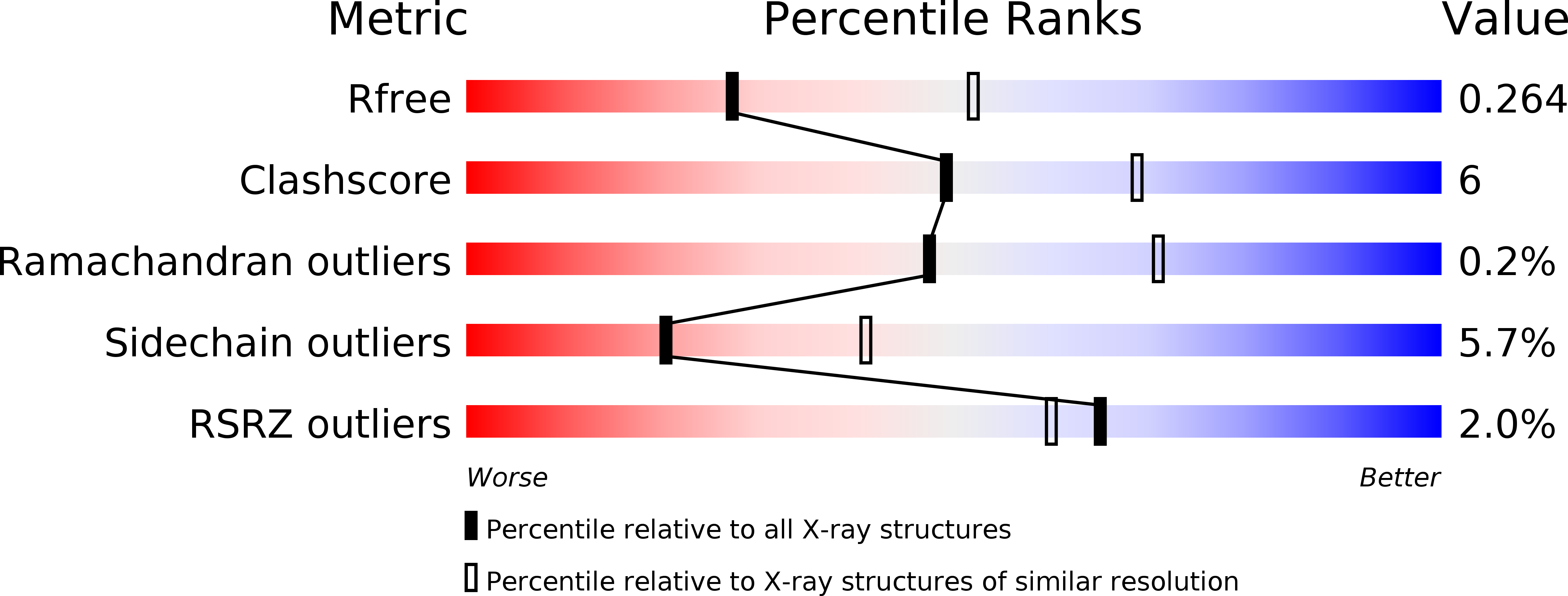
Resolution:
2.60 Å
R-Value Free:
0.26
R-Value Work:
0.19
R-Value Observed:
0.20
Space Group:
P 21 21 21