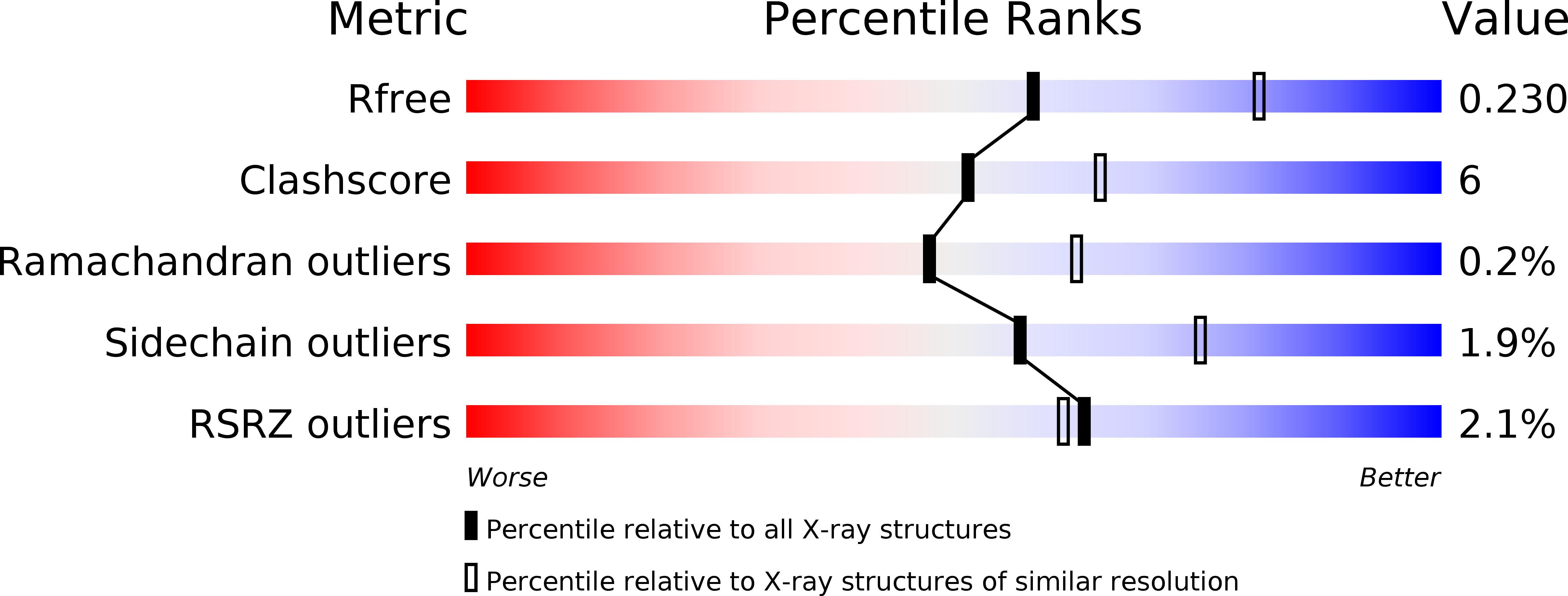
3R0E image
Deposition Date 2011-03-07
Release Date 2011-08-10
Last Version Date 2024-11-20
Entry Detail
PDB ID:
3R0E
Title:
Structure of Remusatia vivipara lectin
Biological Source:
Source Organism:
Method Details:
Experimental Method:
Resolution:
2.40 Å
R-Value Free:
0.23
R-Value Work:
0.20
R-Value Observed:
0.21
Space Group:
P 41
Macromolecular Entities
Structures with similar UniProt ID
Protein Blast
Polymer Type:polypeptide(L)
Molecule:Lectin
Gene (Uniprot):L1
Chain IDs:A, C
Chain Length:109
Number of Molecules:2
Biological Source:Remusatia vivipara
Structures with similar UniProt ID
Protein Blast
Polymer Type:polypeptide(L)
Molecule:Lectin
Gene (Uniprot):L1
Chain IDs:B, D
Chain Length:110
Number of Molecules:2
Biological Source:Remusatia vivipara
Primary Citation
Crystal structure of a {beta}-prism II lectin from Remusatia vivipara.
Glycobiology 22 56 69 (2012)
PMID: 21788359 DOI: 10.1093/glycob/cwr100

Abstact

The crystal structure of a β-prism II (BP2) fold lectin from Remusatia vivipara, a plant of traditional medicinal value, has been determined at a resolution of 2.4 Å. This lectin (RVL, Remusatia vivipara lectin) is a dimer with each protomer having two distinct BP2 domains without a linker between them. It belongs to the "monocot mannose-binding" lectin family, which consists of proteins of high sequence and structural similarity. Though the overall tertiary structure is similar to that of lectins from snowdrop bulbs and garlic, crucial differences in the mannose-binding regions and oligomerization were observed. Unlike most of the other structurally known proteins in this family, only one of the three carbohydrate recognition sites (CRSs) per BP2 domain is found to be conserved. RVL does not recognize simple mannose moieties. RVL binds to only N-linked complex glycans like those present on the gp120 envelope glycoprotein of HIV and mannosylated blood proteins like fetuin, but not to simple mannose moieties. The molecular basis for these features and their possible functional implications to understand the different levels of carbohydrate affinities in this structural family have been investigated through structure analysis, modeling and binding studies. Apart from being the first structure of a lectin to be reported from the Araceae/Arum family, this protein also displays a novel mode of oligomerization among BP2 lectins.

Legend

Protein

Chemical

Disease

Primary Citation of related structures