Deposition Date
2011-03-04
Release Date
2011-03-23
Last Version Date
2024-02-21
Entry Detail
PDB ID:
3QZC
Keywords:
Title:
Structure of the periplasmic stress response protein CpxP
Biological Source:
Source Organism(s):
Escherichia coli (Taxon ID: 83333)
Expression System(s):
Method Details:
Experimental Method:
Resolution:
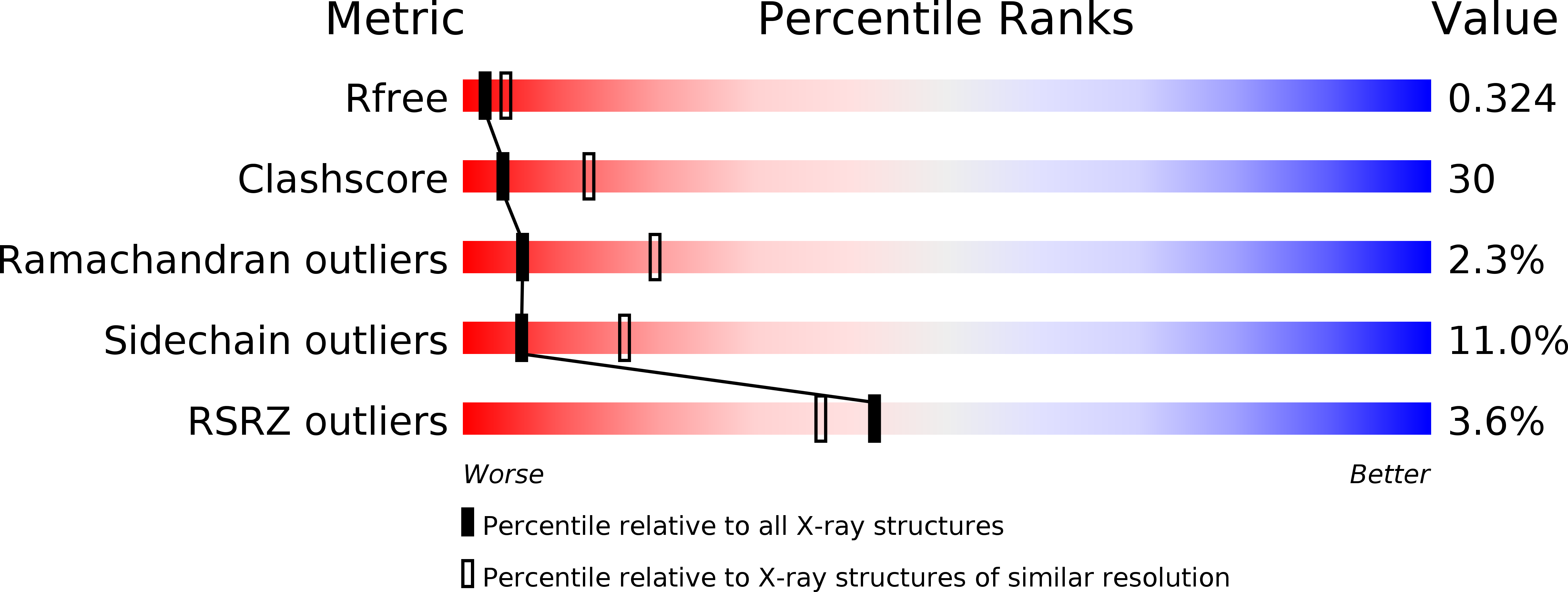
2.85 Å
R-Value Free:
0.29
R-Value Work:
0.24
R-Value Observed:
0.24
Space Group:
P 61 2 2