Deposition Date
2011-03-04
Release Date
2011-08-24
Last Version Date
2025-03-26
Entry Detail
PDB ID:
3QYZ
Keywords:
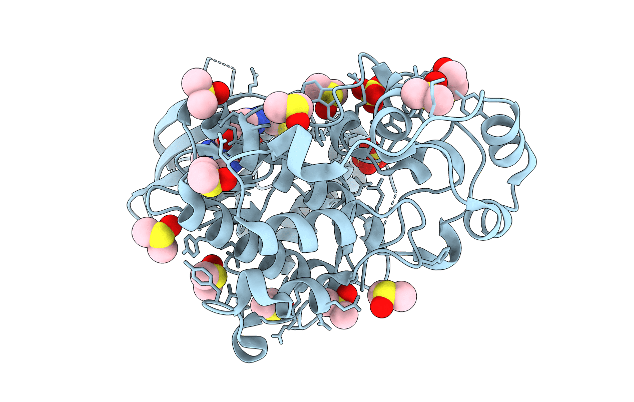
Title:
Crystal structure of ERK2 in complex with an inhibitor
Biological Source:
Source Organism(s):
Rattus norvegicus (Taxon ID: 10116)
Expression System(s):
Method Details:
Experimental Method:
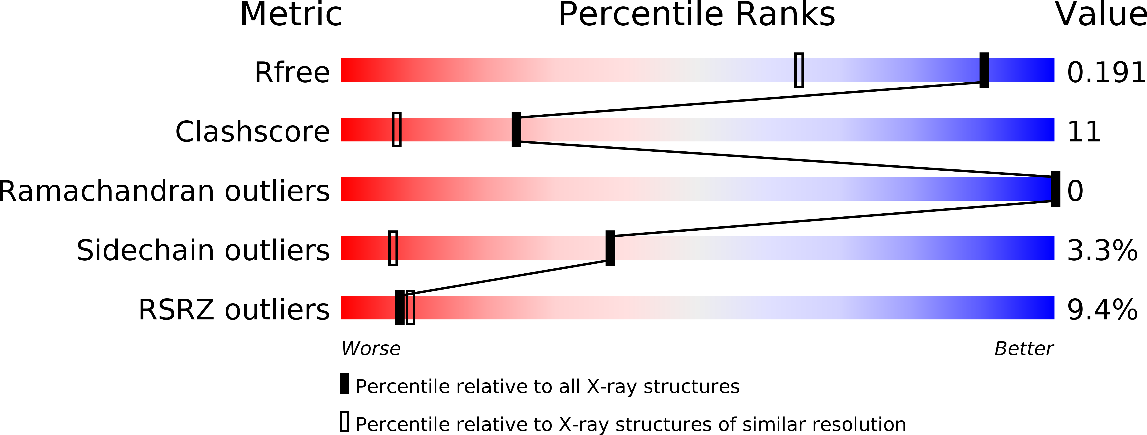
Resolution:
1.46 Å
R-Value Free:
0.19
R-Value Work:
0.16
R-Value Observed:
0.16
Space Group:
P 1 21 1