3QYC image
Deposition Date 2011-03-03
Release Date 2012-02-08
Last Version Date 2024-11-20
Entry Detail
PDB ID:
3QYC
Keywords:
Title:
Structure of a dimeric anti-HER2 single domain antibody
Biological Source:
Source Organism:
Homo sapiens (Taxon ID: 9606)
Host Organism:
Method Details:
Experimental Method:
Resolution:
1.60 Å
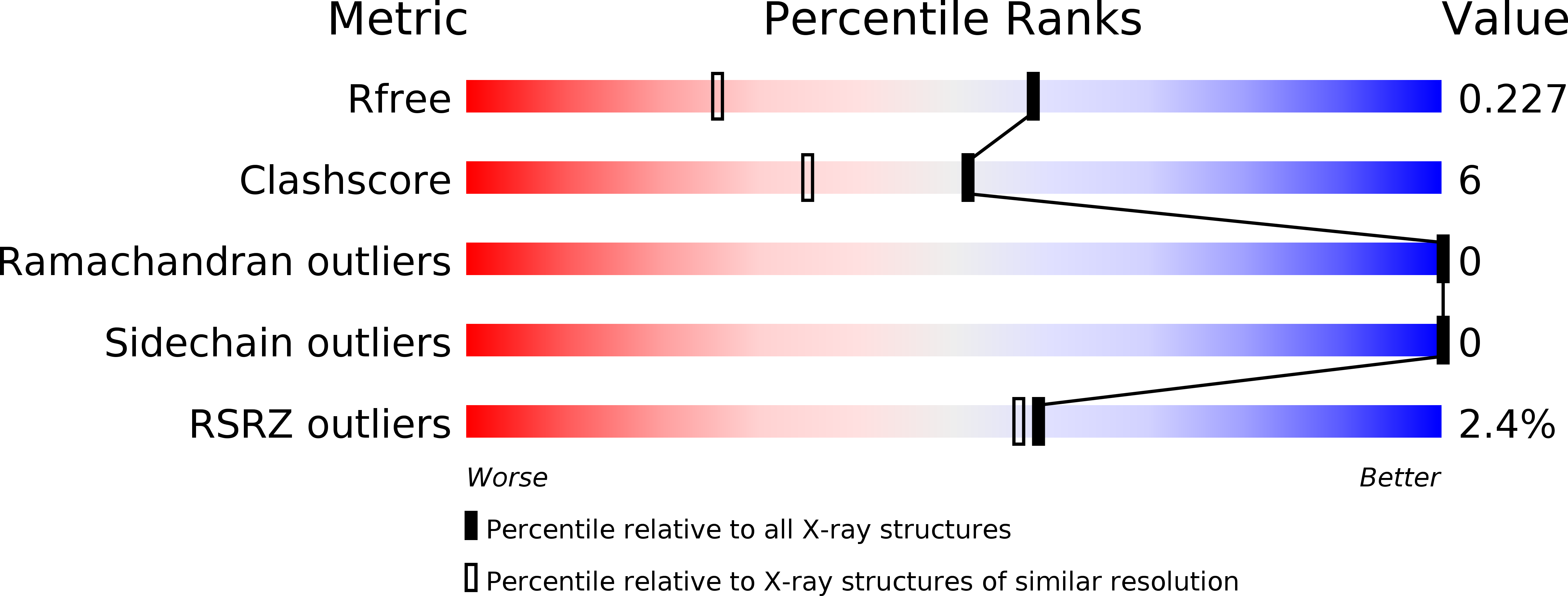
R-Value Free:
0.22
R-Value Work:
0.20
R-Value Observed:
0.20
Space Group:
P 21 21 21
Macromolecular Entities
Polymer Type:polypeptide(L)
Molecule:VH domain of IgG molecule
Chain IDs:A, B
Chain Length:146
Number of Molecules:2
Biological Source:Homo sapiens
Primary Citation
Crystal Structure of a Human Single Domain Antibody Dimer Formed through V(H)-V(H) Non-Covalent Interactions.
Plos One 7 e30149 e30149 (2012)
PMID: 22253912 DOI: 10.1371/journal.pone.0030149

Abstact

Single-domain antibodies (sdAbs) derived from human V(H) are considered to be less soluble and prone to aggregate which makes it difficult to determine the crystal structures. In this study, we isolated and characterized two anti-human epidermal growth factor receptor-2 (HER2) sdAbs, Gr3 and Gr6, from a synthetic human V(H) phage display library. Size exclusion chromatography and surface plasmon resonance analyses demonstrated that Gr3 is a monomer, but that Gr6 is a strict dimer. To understand this different molecular behavior, we solved the crystal structure of Gr6 to 1.6 Å resolution. The crystal structure revealed that the homodimer assembly of Gr6 closely mimics the V(H)-V(L) heterodimer of immunoglobulin variable domains and the dimerization interface is dominated by hydrophobic interactions.

Legend

Protein

Chemical

Disease

Primary Citation of related structures