Deposition Date
2011-02-24
Release Date
2011-11-09
Last Version Date
2024-11-06
Entry Detail
PDB ID:
3QUM
Keywords:
Title:
Crystal structure of human prostate specific antigen (PSA) in Fab sandwich with a high affinity and a PCa selective antibody
Biological Source:
Source Organism(s):
Homo sapiens (Taxon ID: 9606)
Mus musculus (Taxon ID: 10090)
Mus musculus (Taxon ID: 10090)
Method Details:
Experimental Method:
Resolution:
3.20 Å
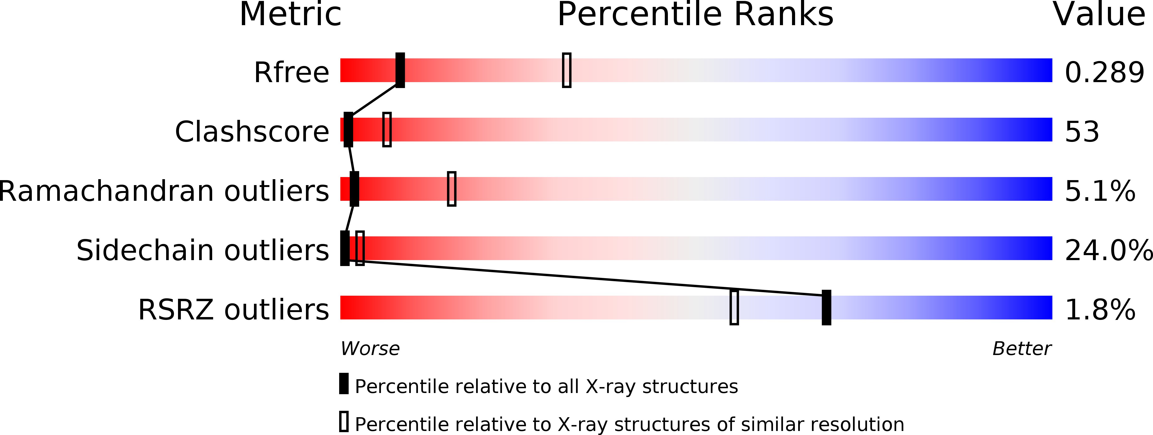
R-Value Free:
0.29
R-Value Work:
0.19
R-Value Observed:
0.20
Space Group:
P 1 21 1