Deposition Date
2011-02-22
Release Date
2012-02-22
Last Version Date
2024-11-06
Entry Detail
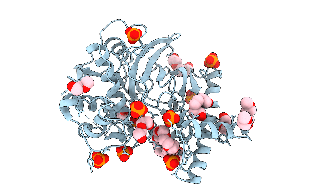
PDB ID:
3QT4
Keywords:
Title:
Structure of digestive procathepsin L 3 of Tenebrio molitor larval midgut
Biological Source:
Source Organism(s):
Tenebrio molitor (Taxon ID: 7067)
Expression System(s):
Method Details:
Experimental Method:
Resolution:
2.11 Å
R-Value Free:
0.20
R-Value Work:
0.15
R-Value Observed:
0.16
Space Group:
C 1 2 1