3QT1 image
Deposition Date 2011-02-22
Release Date 2011-03-23
Last Version Date 2024-11-20
Entry Detail
PDB ID:
3QT1
Title:
RNA polymerase II variant containing A Chimeric RPB9-C11 subunit
Biological Source:
Source Organism:
Host Organism:
Method Details:
Experimental Method:
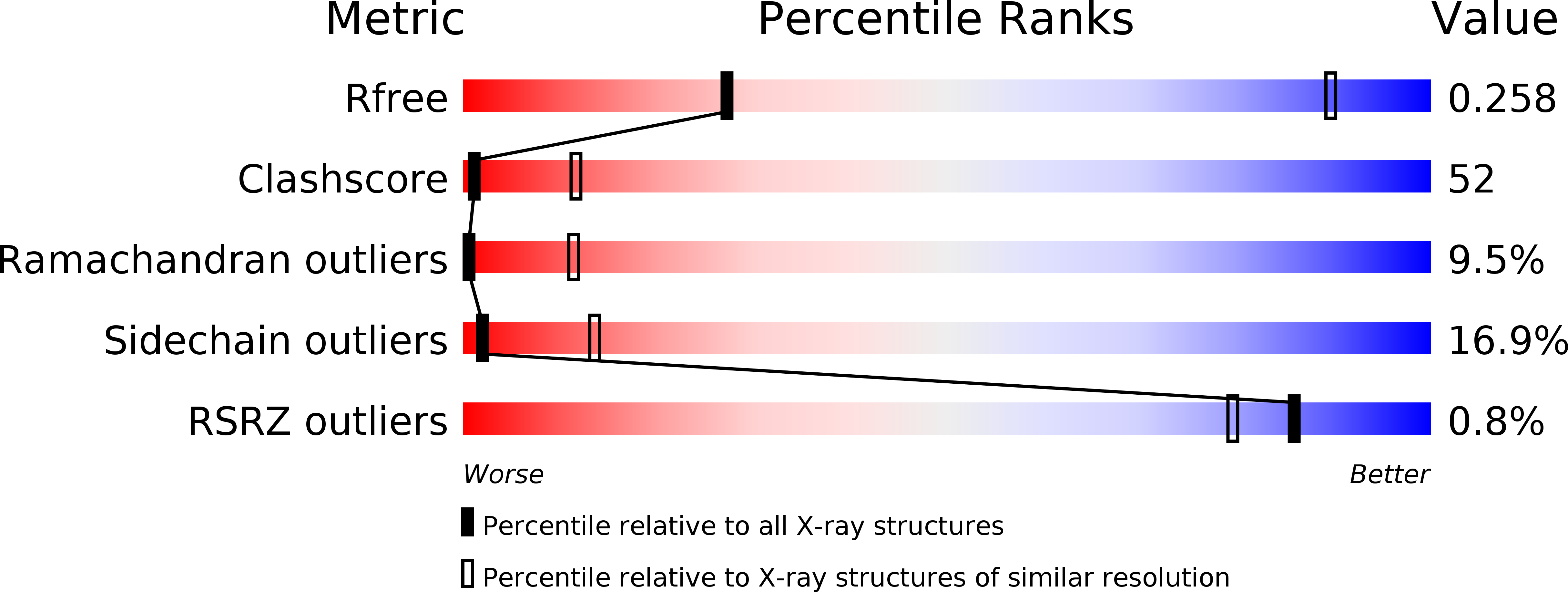
Resolution:
4.30 Å
R-Value Free:
0.28
R-Value Work:
0.23
R-Value Observed:
0.23
Space Group:
C 2 2 21
Macromolecular Entities
Structures with similar UniProt ID
Protein Blast
Polymer Type:polypeptide(L)
Molecule:DNA-directed RNA polymerase II subunit RPB1
Gene (Uniprot):RPO21
Chain IDs:A
Chain Length:1733
Number of Molecules:1
Biological Source:Saccharomyces cerevisiae
Structures with similar UniProt ID
Protein Blast
Polymer Type:polypeptide(L)
Molecule:DNA-directed RNA polymerase II subunit RPB2
Gene (Uniprot):RPB2
Chain IDs:B
Chain Length:1224
Number of Molecules:1
Biological Source:Saccharomyces cerevisiae
Structures with similar UniProt ID
Protein Blast
Polymer Type:polypeptide(L)
Molecule:DNA-directed RNA polymerase II subunit RPB3
Gene (Uniprot):RPB3
Chain IDs:C
Chain Length:318
Number of Molecules:1
Biological Source:Saccharomyces cerevisiae
Structures with similar UniProt ID
Protein Blast
Polymer Type:polypeptide(L)
Molecule:DNA-directed RNA polymerase II subunit RPB4
Gene (Uniprot):RPB4
Chain IDs:D
Chain Length:219
Number of Molecules:1
Biological Source:Saccharomyces cerevisiae
Structures with similar UniProt ID
Protein Blast
Polymer Type:polypeptide(L)
Molecule:DNA-directed RNA polymerases I, II, and III subunit RPABC1
Gene (Uniprot):RPB5
Chain IDs:E
Chain Length:215
Number of Molecules:1
Biological Source:Saccharomyces cerevisiae
Structures with similar UniProt ID
Protein Blast
Polymer Type:polypeptide(L)
Molecule:DNA-directed RNA polymerases I, II, and III subunit RPABC2
Gene (Uniprot):RPO26
Chain IDs:F
Chain Length:155
Number of Molecules:1
Biological Source:Saccharomyces cerevisiae
Structures with similar UniProt ID
Protein Blast
Polymer Type:polypeptide(L)
Molecule:DNA-directed RNA polymerase II subunit RPB7
Gene (Uniprot):RPB7
Chain IDs:G
Chain Length:171
Number of Molecules:1
Biological Source:Saccharomyces cerevisiae
Structures with similar UniProt ID
Protein Blast
Polymer Type:polypeptide(L)
Molecule:DNA-directed RNA polymerases I, II, and III subunit RPABC3
Gene (Uniprot):RPB8
Chain IDs:H
Chain Length:146
Number of Molecules:1
Biological Source:Saccharomyces cerevisiae
Protein Blast
Polymer Type:polypeptide(L)
Molecule:DNA-directed RNA polymerase II subunit RPB9, DNA-directed RNA polymerase III subunit RPC10
Gene (Uniprot):RPB9, RPC11
Chain IDs:I
Chain Length:133
Number of Molecules:1
Biological Source:Saccharomyces cerevisiae
Structures with similar UniProt ID
Protein Blast
Polymer Type:polypeptide(L)
Molecule:DNA-directed RNA polymerases I, II, and III subunit RPABC5
Gene (Uniprot):RPB10
Chain IDs:J
Chain Length:70
Number of Molecules:1
Biological Source:Saccharomyces cerevisiae
Structures with similar UniProt ID
Protein Blast
Polymer Type:polypeptide(L)
Molecule:DNA-directed RNA polymerase II subunit RPB11
Gene (Uniprot):RPB11
Chain IDs:K
Chain Length:120
Number of Molecules:1
Biological Source:Saccharomyces cerevisiae
Structures with similar UniProt ID
Protein Blast
Polymer Type:polypeptide(L)
Molecule:DNA-directed RNA polymerases I, II, and III subunit RPABC4
Gene (Uniprot):RPC10
Chain IDs:L
Chain Length:70
Number of Molecules:1
Biological Source:Saccharomyces cerevisiae
Primary Citation
Evolution of two modes of intrinsic RNA polymerase transcript cleavage.
J.Biol.Chem. 286 18701 18707 (2011)
PMID: 21454497 DOI: 10.1074/jbc.M111.222273

Abstact

During gene transcription, the RNA polymerase (Pol) active center can catalyze RNA cleavage. This intrinsic cleavage activity is strong for Pol I and Pol III but very weak for Pol II. The reason for this difference is unclear because the active centers of the polymerases are virtually identical. Here we show that Pol II gains strong cleavage activity when the C-terminal zinc ribbon domain (C-ribbon) of subunit Rpb9 is replaced by its counterpart from the Pol III subunit C11. X-ray analysis shows that the C-ribbon has detached from its site on the Pol II surface and is mobile. Mutagenesis indicates that the C-ribbon transiently inserts into the Pol II pore to complement the active center. This mechanism is also used by transcription factor IIS, a factor that can bind Pol II and induce strong RNA cleavage. Together with published data, our results indicate that Pol I and Pol III contain catalytic C-ribbons that complement the active center, whereas Pol II contains a non-catalytic C-ribbon that is immobilized on the enzyme surface. Evolution of the Pol II system may have rendered mRNA transcript cleavage controllable by the dissociable factor transcription factor IIS to enable promoter-proximal gene regulation and elaborate 3'-processing and transcription termination.

Legend

Protein

Chemical

Disease

Primary Citation of related structures
Feedback Form
Name
Email
Institute
Feedback