Deposition Date
2011-02-21
Release Date
2012-04-04
Last Version Date
2024-02-21
Entry Detail
PDB ID:
3QSI
Keywords:
Title:
Nickel binding domain of NikR from Helicobacter pylori disclosing partial metal occupancy
Biological Source:
Source Organism(s):
Helicobacter pylori (Taxon ID: 210)
Expression System(s):
Method Details:
Experimental Method:
Resolution:
3.08 Å
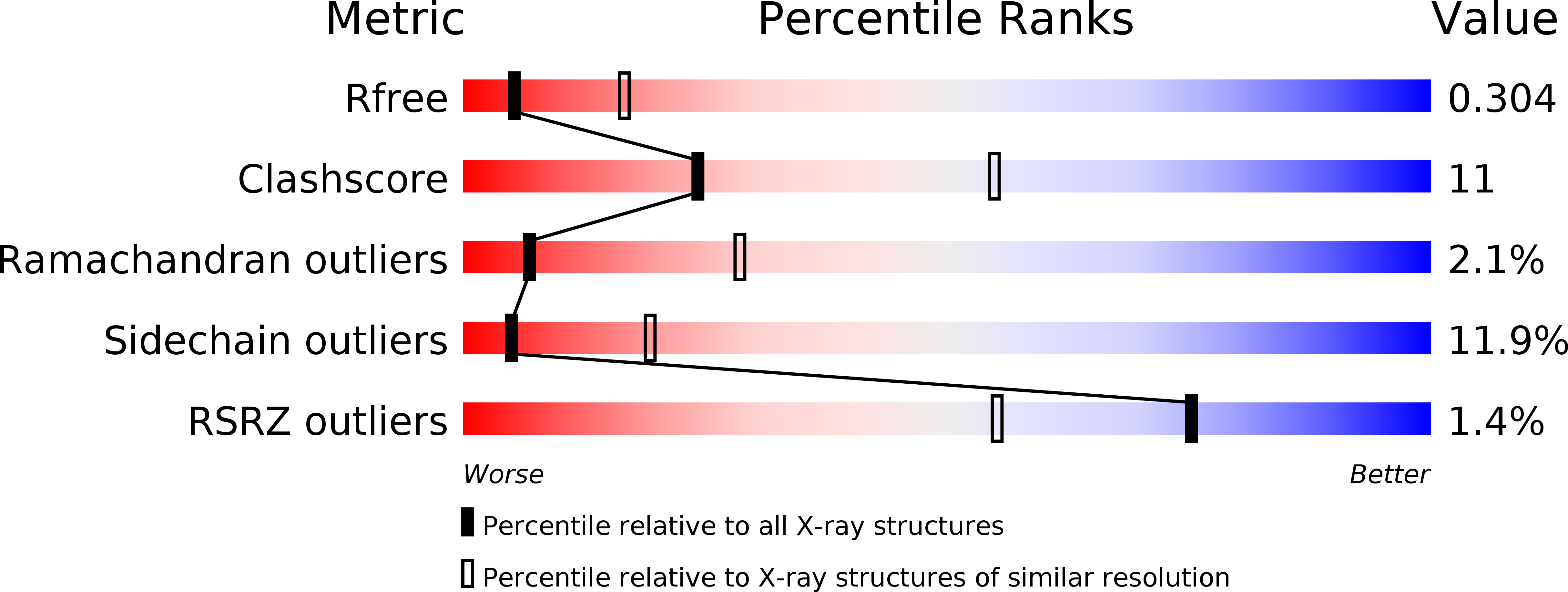
R-Value Free:
0.30
R-Value Work:
0.25
R-Value Observed:
0.26
Space Group:
C 1 2 1