3QRX image
Deposition Date 2011-02-18
Release Date 2011-10-26
Last Version Date 2023-09-13
Entry Detail
PDB ID:
3QRX
Title:
Chlamydomonas reinhardtii centrin bound to melittin
Biological Source:
Source Organism:
Host Organism:
Method Details:
Experimental Method:
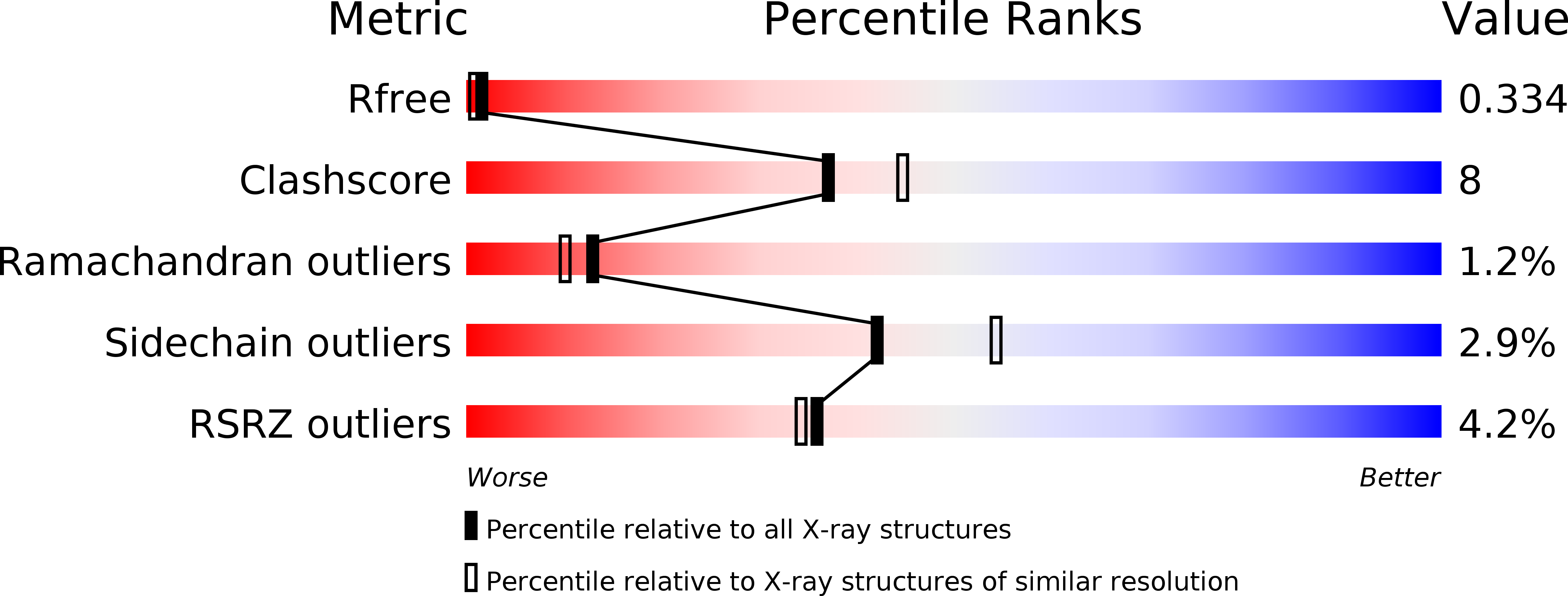
Resolution:
2.20 Å
R-Value Free:
0.34
R-Value Work:
0.29
R-Value Observed:
0.29
Space Group:
P 21 21 2
Macromolecular Entities
Structures with similar UniProt ID
Protein Blast
Polymer Type:polypeptide(L)
Molecule:Centrin
Chain IDs:A
Chain Length:169
Number of Molecules:1
Biological Source:Chlamydomonas reinhardtii
Structures with similar UniProt ID
Protein Blast
Polymer Type:polypeptide(L)
Molecule:Melittin
Gene (Uniprot):MELT
Chain IDs:B
Chain Length:26
Number of Molecules:1
Biological Source:
Ligand Molecules
Primary Citation
The structure, molecular dynamics, and energetics of centrin-melittin complex.
Proteins 79 3132 3143 (2011)
PMID: 21989934 DOI: 10.1002/prot.23142

Abstact

Centrin is a calcium binding protein (CaBP) belonging to the EF-hand superfamily. As with other proteins within this family, centrin is a calcium sensor with multiple biological target proteins. We chose to study Chlamydomonas reinhardtii centrin (Crcen) and its interaction with melittin (MLT) as a model for CaBP complexes due to its amphipathic properties. Our goal was to determine the molecular interactions that lead to centrin-MLT complex formation, their relative stability, and the conformational changes associated with the interaction, when compared to the single components. For this, we determined the thermodynamic parameters that define Crcen-MLT complex formation. Two-dimensional infrared (2D IR) correlation spectroscopy were used to study the amide I', I'*, and side chain bands for (13)C-Crcen, MLT, and the (13)C-Crcen-MLT complex. This approach resulted in the determination of MLT's increased helicity, while centrin was stabilized within the complex. Herein we provide the first complete molecular description of centrin-MLT complex formation and the dissociation process. Also, discussed is the first structure of a CaBP-MLT complex by X-ray crystallography, which shows that MLT has a different binding orientation than previously characterized centrin-bound peptides. Finally, all of the experimental results presented herein are consistent with centrin maintaining an extended conformation while interacting with MLT. The molecular implications of these results are: (1) the recognition of hydrophobic contacts as requirements for initial binding, (2) minimum electrostatic interactions within the C-terminal end of the peptide, and (3) van der Waals interactions within MLTs N-terminal end are required for complex formation.

Legend

Protein

Chemical

Disease

Primary Citation of related structures
Feedback Form
Name
Email
Institute
Feedback