
Deposition Date
2011-02-17
Release Date
2012-02-22
Last Version Date
2024-10-09
Entry Detail
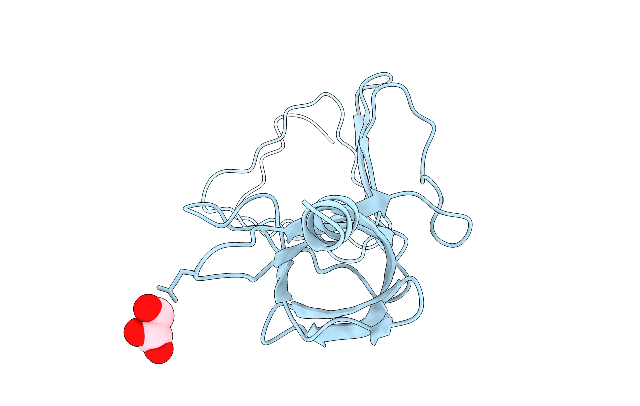
PDB ID:
3QR8
Keywords:
Title:
Crystal structure of the bacteriophage P2 membrane-piercing protein gpV
Biological Source:
Source Organism(s):
Enterobacteria phage P2 (Taxon ID: 10679)
Expression System(s):
Method Details:
Experimental Method:
Resolution:
2.03 Å
R-Value Free:
0.30
R-Value Work:
0.25
R-Value Observed:
0.26
Space Group:
P 3 2 1
