3QR5 image
Deposition Date 2011-02-17
Release Date 2011-06-08
Last Version Date 2024-11-27
Entry Detail
PDB ID:
3QR5
Title:
Structure of the first domain of a cardiac Ryanodine Receptor mutant with exon 3 deleted
Biological Source:
Source Organism:
Mus musculus (Taxon ID: 10090)
Host Organism:
Method Details:
Experimental Method:
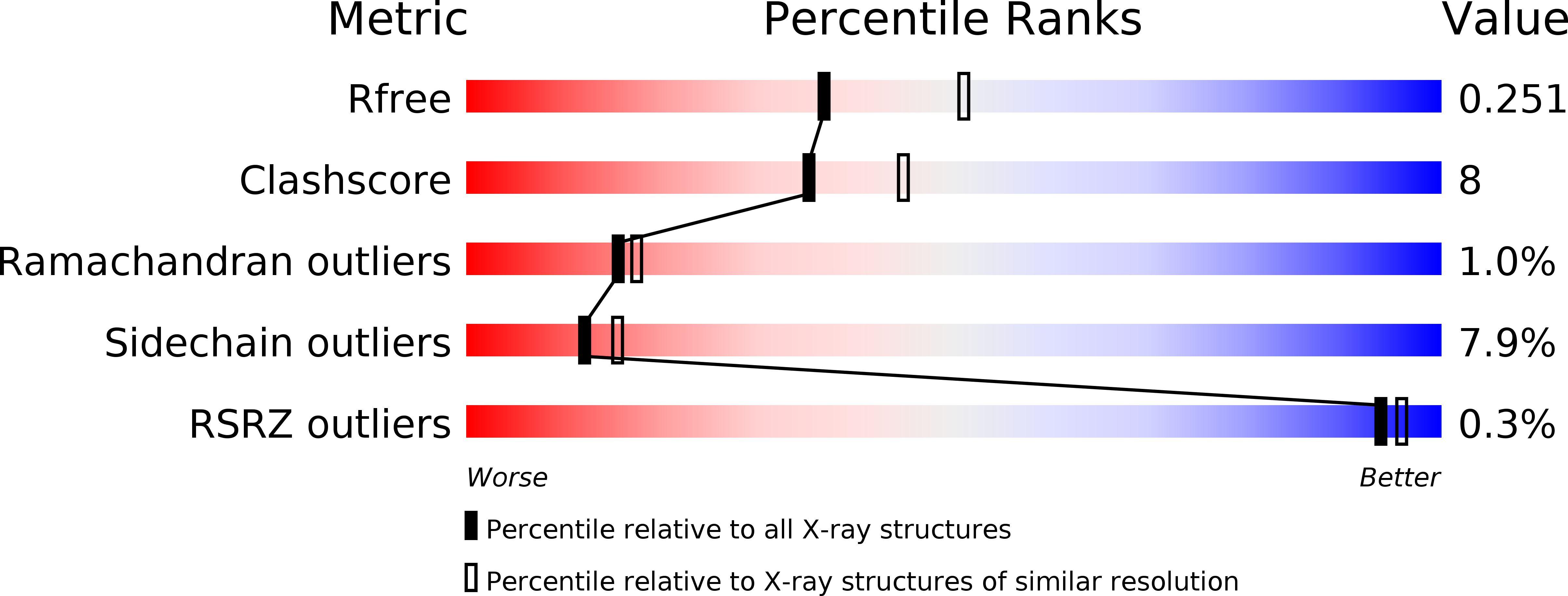
Resolution:
2.30 Å
R-Value Free:
0.25
R-Value Work:
0.20
R-Value Observed:
0.21
Space Group:
C 1 2 1
Macromolecular Entities
Polymer Type:polypeptide(L)
Molecule:Cardiac Ca2+ release channel
Chain IDs:A, B
Chain Length:185
Number of Molecules:2
Biological Source:Mus musculus
Primary Citation
The deletion of exon 3 in the cardiac ryanodine receptor is rescued by beta strand switching.
Structure 19 790 798 (2011)
PMID: 21645850 DOI: 10.1016/j.str.2011.03.016

Abstact

Mutations in the cardiac Ryanodine Receptor (RYR2) are linked to triggered arrhythmias. Removal of exon 3 results in a severe form of catecholaminergic polymorphic ventricular tachycardia (CPVT). This exon encodes secondary structure elements that are crucial for folding of the N-terminal domain (NTD), raising the question of why the deletion is neither lethal nor confers a loss of function. We determined the 2.3 Å crystal structure of the NTD lacking exon 3. The removal causes a structural rescue whereby a flexible loop inserts itself into the β trefoil domain and increases thermal stability. The exon 3 deletion is not tolerated in the corresponding RYR1 domain. The rescue shows a novel mechanism by which RYR2 channels can adjust their Ca²⁺ release properties through altering the structure of the NTD. Despite the rescue, the deletion affects interfaces with other RYR2 domains. We propose that relative movement of the NTD is allosterically coupled to the pore region.

Legend

Protein

Chemical

Disease

Primary Citation of related structures