3QQ5 image
Deposition Date 2011-02-15
Release Date 2011-11-16
Last Version Date 2024-11-06
Entry Detail
PDB ID:
3QQ5
Keywords:
Title:
Crystal structure of the [FeFe]-hydrogenase maturation protein HydF
Biological Source:
Source Organism:
Host Organism:
Method Details:
Experimental Method:
Resolution:
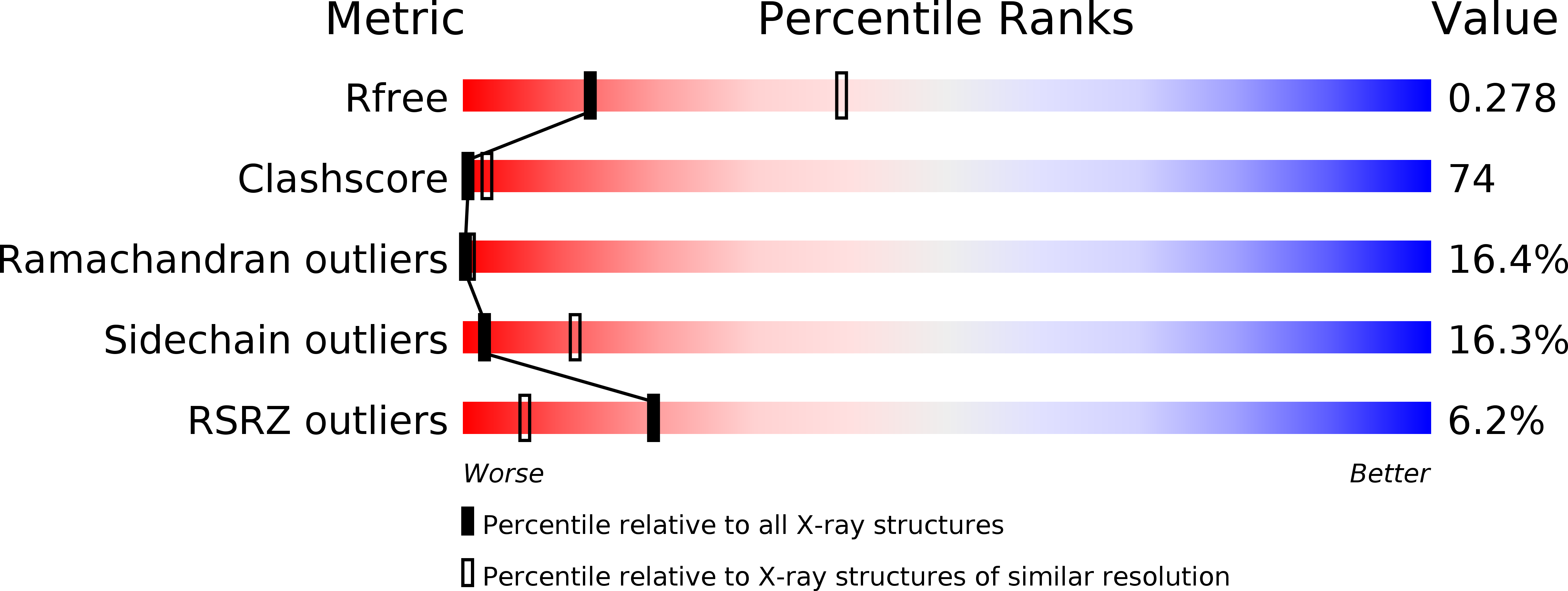
2.99 Å
R-Value Free:
0.30
R-Value Work:
0.27
R-Value Observed:
0.27
Space Group:
P 2 3
Macromolecular Entities
Polymer Type:polypeptide(L)
Molecule:Small GTP-binding protein
Gene (Uniprot):CTN_0227
Chain IDs:A
Chain Length:423
Number of Molecules:1
Biological Source:Thermotoga neapolitana
Ligand Molecules
Primary Citation
Crystal Structure of HydF Scaffold Protein Provides Insights into [FeFe]-Hydrogenase Maturation.
J.Biol.Chem. 286 43944 43950 (2011)
PMID: 22057316 DOI: 10.1074/jbc.M111.281956

Abstact

[FeFe]-hydrogenases catalyze the reversible production of H2 in some bacteria and unicellular eukaryotes. These enzymes require ancillary proteins to assemble the unique active site H-cluster, a complex structure composed of a 2Fe center bridged to a [4Fe-4S] cubane. The first crystal structure of a key factor in the maturation process, HydF, has been determined at 3 Å resolution. The protein monomer present in the asymmetric unit of the crystal comprises three domains: a GTP-binding domain, a dimerization domain, and a metal cluster-binding domain, all characterized by similar folding motifs. Two monomers dimerize, giving rise to a stable dimer, held together mainly by the formation of a continuous β-sheet comprising eight β-strands from two monomers. Moreover, in the structure presented, two dimers aggregate to form a supramolecular organization that represents an inactivated form of the HydF maturase. The crystal structure of the latter furnishes several clues about the events necessary for cluster generation/transfer and provides an excellent model to begin elucidating the structure/function of HydF in [FeFe]-hydrogenase maturation.

Legend

Protein

Chemical

Disease

Primary Citation of related structures