3QPH image
Deposition Date 2011-02-13
Release Date 2012-05-30
Last Version Date 2024-02-21
Entry Detail
PDB ID:
3QPH
Keywords:
Title:
The three-dimensional structure of TrmB, a global transcriptional regulator of the hyperthermophilic archaeon Pyrococcus furiosus in complex with sucrose
Biological Source:
Source Organism:
Host Organism:
Method Details:
Experimental Method:
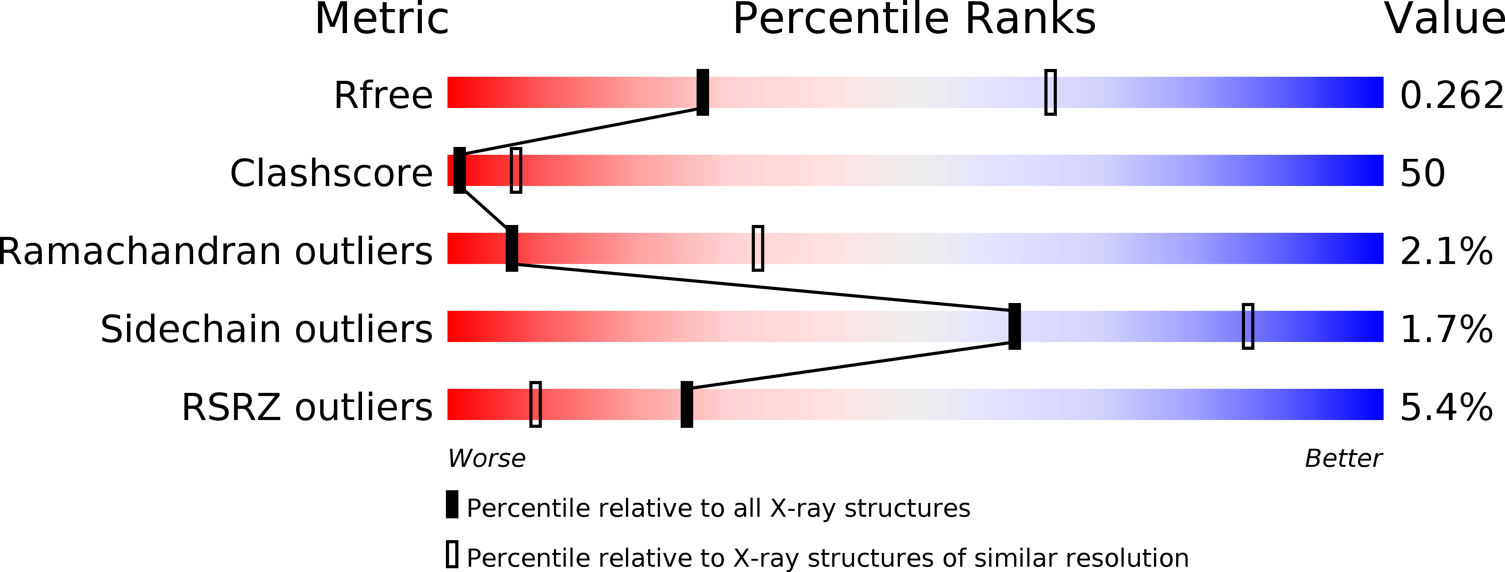
Resolution:
2.99 Å
R-Value Free:
0.26
R-Value Work:
0.22
R-Value Observed:
0.22
Space Group:
P 32 2 1
Macromolecular Entities
Structures with similar UniProt ID
Protein Blast
Polymer Type:polypeptide(L)
Molecule:TrmB, a global transcription regulator
Gene (Uniprot):trmB
Chain IDs:A
Chain Length:342
Number of Molecules:1
Biological Source:Pyrococcus furiosus
Peptide-like Molecules
PRD_900003
Primary Citation
The three-dimensional structure of TrmB, a transcriptional regulator of dual function in the hyperthermophilic archaeon Pyrococcus furiosus in complex with sucrose.
Protein Sci. 22 800 808 (2013)
PMID: 23576322 DOI: 10.1002/pro.2263

Abstact

TrmB is a repressor that binds maltose, maltotriose, and sucrose, as well as other α-glucosides. It recognizes two different operator sequences controlling the TM (Trehalose/Maltose) and the MD (Maltodextrin) operon encoding the respective ABC transporters and sugar-degrading enzymes. Binding of maltose to TrmB abrogates repression of the TM operon but maintains the repression of the MD operon. On the other hand, binding of sucrose abrogates repression of the MD operon but maintains repression of the TM operon. The three-dimensional structure of TrmB in complex with sucrose was solved and refined to a resolution of 3.0 Å. The structure shows the N-terminal DNA binding domain containing a winged-helix-turn-helix (wHTH) domain followed by an amphipathic helix with a coiled-coil motif. The latter promotes dimerization and places the symmetry mates of the putative recognition helix in the wHTH motif about 30 Å apart suggesting a canonical binding to two successive major grooves of duplex palindromic DNA. This suggests that the structure resembles the conformation of TrmB recognizing the pseudopalindromic TM promoter but not the conformation recognizing the nonpalindromic MD promoter.

Legend

Protein

Chemical

Disease

Primary Citation of related structures