Deposition Date
2011-02-11
Release Date
2011-03-30
Last Version Date
2023-09-13
Entry Detail
PDB ID:
3QP8
Keywords:
Title:
Crystal structure of CviR (Chromobacterium violaceum 12472) ligand-binding domain bound to C10-HSL
Biological Source:
Source Organism(s):
Chromobacterium violaceum (Taxon ID: 536)
Expression System(s):
Method Details:
Experimental Method:
Resolution:
1.60 Å
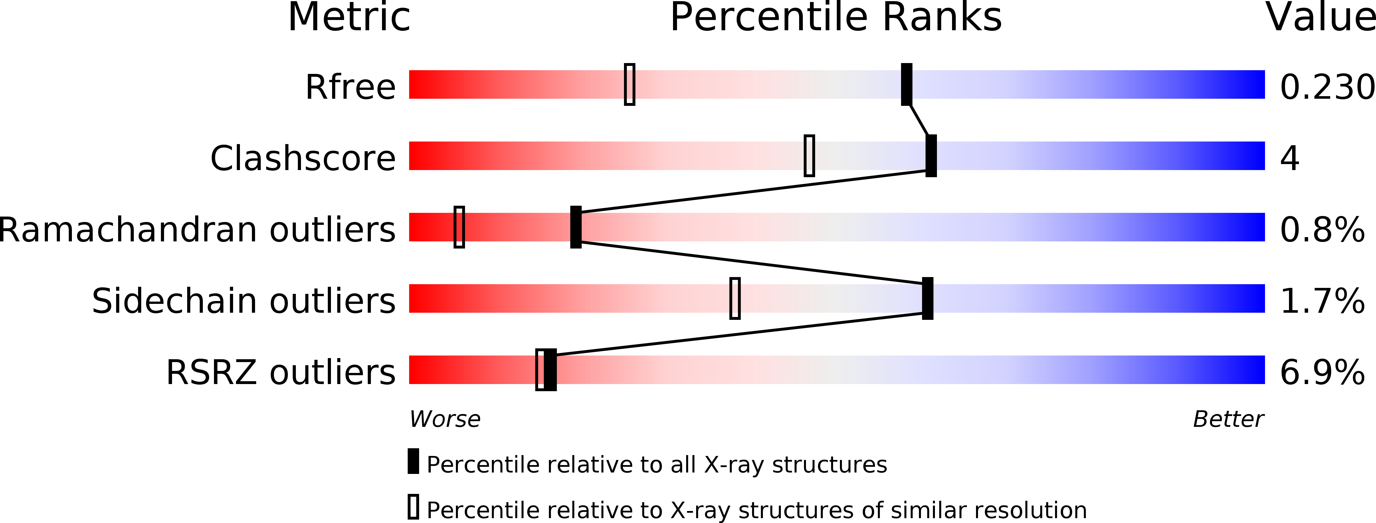
R-Value Free:
0.23
R-Value Work:
0.19
R-Value Observed:
0.19
Space Group:
P 1 21 1