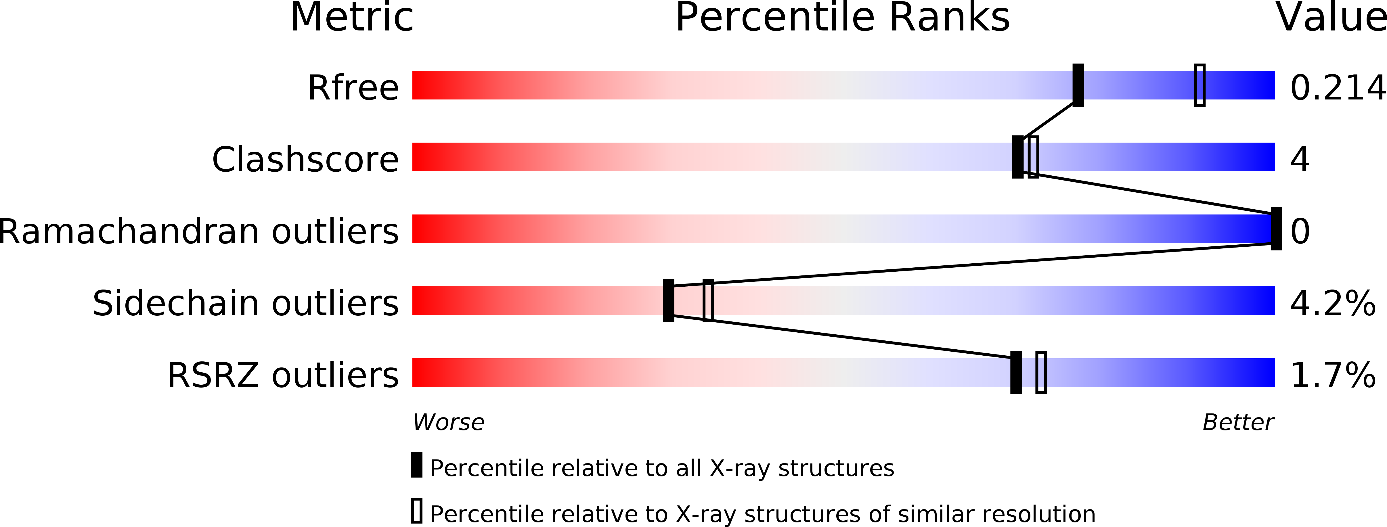
3QNR image
Entry Detail
PDB ID:
3QNR
Keywords:
Title:
DyPB from Rhodococcus jostii RHA1, crystal form 1
Biological Source:
Source Organism:
Host Organism:
PDB Version:
Deposition Date:
2011-02-09
Release Date:
2011-04-27
Method Details:
Experimental Method:
Resolution:
2.25 Å
R-Value Free:
0.21
R-Value Work:
0.19
R-Value Observed:
0.19
Space Group:
P 32 2 1
Macromolecular Entities
Polymer Type:polypeptide(L)
Description:DyP Peroxidase
Chain IDs:A, B, C
Chain Length:353
Number of Molecules:3
Biological Source:Rhodococcus jostii RHA1
Primary Citation
Characterization of dye-decolorizing peroxidases from Rhodococcus jostii RHA1.
Biochemistry 50 5108 5119 (2011)
PMID: 21534572 DOI: 10.1021/bi200427h

Abstact

The soil bacterium Rhodococcus jostii RHA1 contains two dye-decolorizing peroxidases (DyPs) named according to the subfamily they represent: DypA, predicted to be periplasmic, and DypB, implicated in lignin degradation. Steady-state kinetic studies of these enzymes revealed that they have much lower peroxidase activities than C- and D-type DyPs. Nevertheless, DypA showed 6-fold greater apparent specificity for the anthraquinone dye Reactive Blue 4 (k(cat)/K(m) = 12800 ± 600 M(-1) s(-1)) than either ABTS or pyrogallol, consistent with previously characterized DyPs. By contrast, DypB showed the greatest apparent specificity for ABTS (k(cat)/K(m) = 2000 ± 100 M(-1) s(-1)) and also oxidized Mn(II) (k(cat)/K(m) = 25.1 ± 0.1 M(-1) s(-1)). Further differences were detected using electron paramagnetic resonance (EPR) spectroscopy: while both DyPs contained high-spin (S = (5)/(2)) Fe(III) in the resting state, DypA had a rhombic high-spin signal (g(y) = 6.32, g(x) = 5.45, and g(z) = 1.97) while DypB had a predominantly axial signal (g(y) = 6.09, g(x) = 5.45, and g(z) = 1.99). Moreover, DypA reacted with H(2)O(2) to generate an intermediate with features of compound II (Fe(IV)═O). By contrast, DypB reacted with H(2)O(2) with a second-order rate constant of (1.79 ± 0.06) × 10(5) M(-1) s(-1) to generate a relatively stable green-colored intermediate (t(1/2) ∼ 9 min). While the electron absorption spectrum of this intermediate was similar to that of compound I of plant-type peroxidases, its EPR spectrum was more consistent with a poorly coupled protein-based radical than with an [Fe(IV)═O Por(•)](+) species. The X-ray crystal structure of DypB, determined to 1.4 Å resolution, revealed a hexacoordinated heme iron with histidine and a solvent species occupying axial positions. A solvent channel potentially provides access to the distal face of the heme for H(2)O(2). A shallow pocket exposes heme propionates to the solvent and contains a cluster of acidic residues that potentially bind Mn(II). Insight into the structure and function of DypB facilitates its engineering for the improved degradation of lignocellulose.

Legend

Protein

Chemical

Disease

Primary Citation of related structures