3QN2 image
Deposition Date 2011-02-07
Release Date 2011-06-01
Last Version Date 2024-11-27
Entry Detail
PDB ID:
3QN2
Keywords:
Title:
Structure-based design of a disulfide-linked oligomeric form of the Simian Virus 40 (SV40) large T antigen DNA binding domain
Biological Source:
Source Organism:
Host Organism:
Method Details:
Experimental Method:
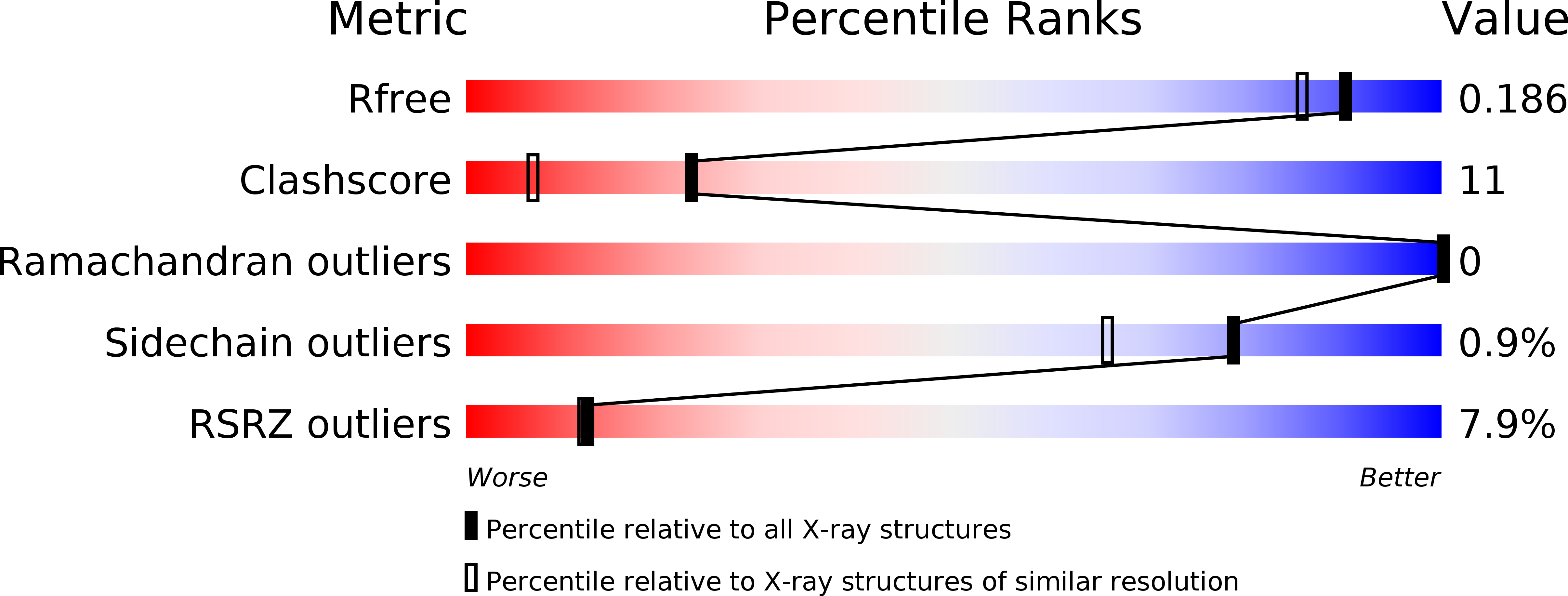
Resolution:
1.66 Å
R-Value Free:
0.19
R-Value Work:
0.16
R-Value Observed:
0.17
Space Group:
P 65
Macromolecular Entities
Polymer Type:polypeptide(L)
Molecule:Large T antigen
Mutations:F151C, C216A, D256C
Chain IDs:A
Chain Length:134
Number of Molecules:1
Biological Source:Simian virus 40
Ligand Molecules
Primary Citation
Structure-based design of a disulfide-linked oligomeric form of the simian virus 40 (SV40) large T antigen DNA-binding domain.
Acta Crystallogr.,Sect.D 67 560 567 (2011)
PMID: 21636896 DOI: 10.1107/S0907444911014302

Abstact

The modular multifunctional protein large T antigen (T-ag) from simian virus 40 orchestrates many of the events needed for replication of the viral double-stranded DNA genome. This protein assembles into single and double hexamers on specific DNA sequences located at the origin of replication. This complicated process begins when the origin-binding domain of large T antigen (T-ag ODB) binds the GAGGC sequences in the central region (site II) of the viral origin of replication. While many of the functions of purified T-ag OBD can be studied in isolation, it is primarily monomeric in solution and cannot assemble into hexamers. To overcome this limitation, the possibility of engineering intermolecular disulfide bonds in the origin-binding domain which could oligomerize in solution was investigated. A recent crystal structure of the wild-type T-ag OBD showed that this domain forms a left-handed spiral in the crystal with six subunits per turn. Therefore, we analyzed the protein interface of this structure and identified two residues that could potentially support an intermolecular disulfide bond if changed to cysteines. SDS-PAGE analysis established that the mutant T-ag OBD formed higher oligomeric products in a redox-dependent manner. In addition, the 1.7 Å resolution crystal structure of the engineered disulfide-linked T-ag OBD is reported, which establishes that oligomerization took place in the expected manner.

Legend

Protein

Chemical

Disease

Primary Citation of related structures