Deposition Date
2011-02-03
Release Date
2011-10-19
Last Version Date
2024-11-06
Entry Detail
PDB ID:
3QLP
Keywords:
Title:
X-ray structure of the complex between human alpha thrombin and a modified thrombin binding aptamer (mTBA)
Biological Source:
Source Organism(s):
Homo sapiens (Taxon ID: 9606)
Expression System(s):
Method Details:
Experimental Method:
Resolution:
2.14 Å
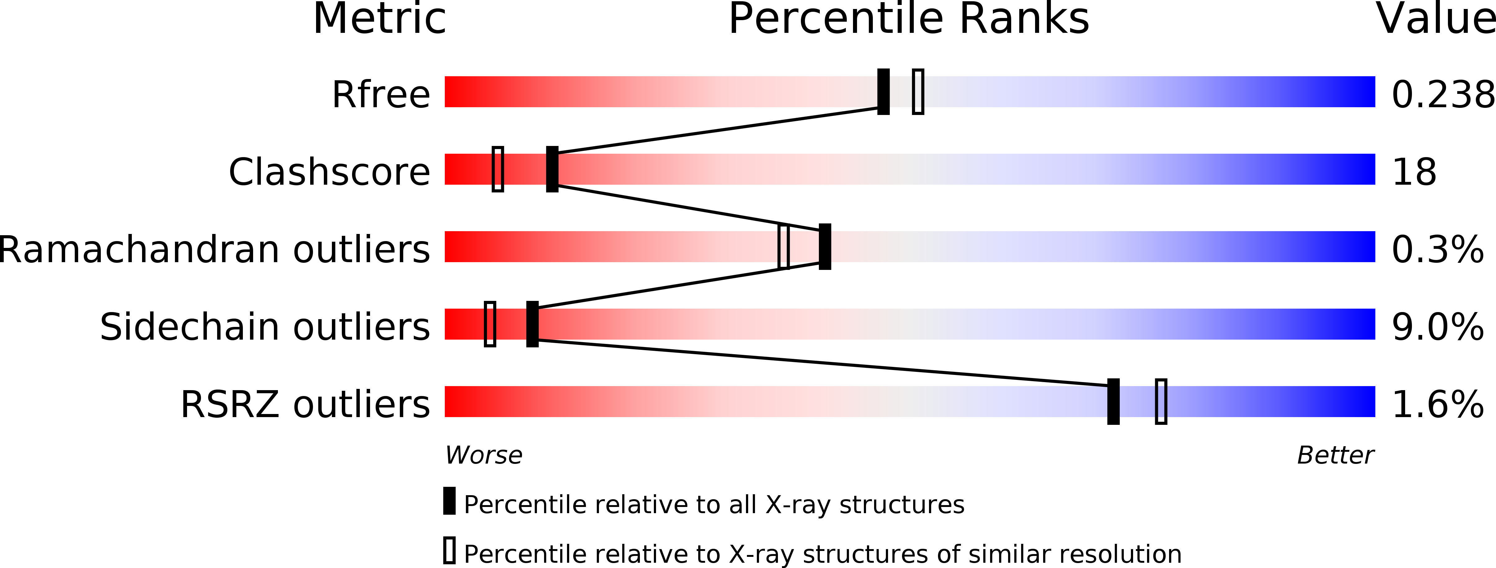
R-Value Free:
0.23
R-Value Work:
0.18
R-Value Observed:
0.18
Space Group:
I 2 2 2