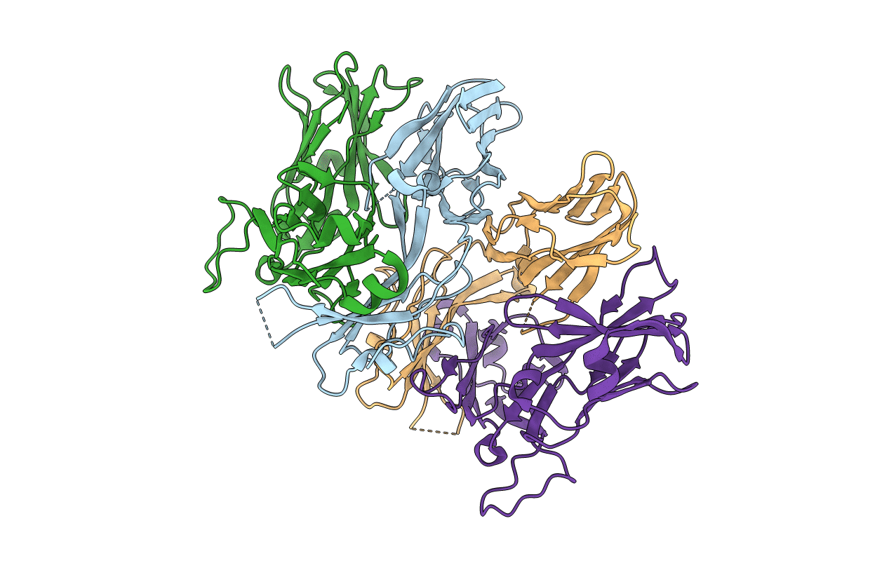
3QJH image
Entry Detail
PDB ID:
3QJH
Keywords:
Title:
The crystal structure of the 5c.c7 TCR
Biological Source:
Source Organism:
Host Organism:
PDB Version:
Deposition Date:
2011-01-28
Release Date:
2011-04-27
Method Details:
Experimental Method:
Resolution:
1.90 Å
R-Value Free:
0.25
R-Value Work:
0.22
R-Value Observed:
0.22
Space Group:
P 21 21 2
Macromolecular Entities
Polymer Type:polypeptide(L)
Description:5c.c7 alpha chain
Chain IDs:A, C
Chain Length:205
Number of Molecules:2
Biological Source:Mus musculus
Polymer Type:polypeptide(L)
Description:5c.c7 beta chain
Chain IDs:B, D
Chain Length:243
Number of Molecules:2
Biological Source:Mus musculus
Primary Citation
Structural basis of specificity and cross-reactivity in T cell receptors specific for cytochrome c-I-E(k).
J.Immunol. 186 5823 5832 (2011)
PMID: 21490152 DOI: 10.4049/jimmunol.1100197

Abstact

T cells specific for the cytochrome c Ag are widely used to investigate many aspects of TCR specificity and interactions with peptide-MHC, but structural information has long been elusive. In this study, we present structures for the well-studied 2B4 TCR, as well as a naturally occurring variant of the 5c.c7 TCR, 226, which is cross-reactive with more than half of possible substitutions at all three TCR-sensitive residues on the peptide Ag. These structures alone and in complex with peptide-MHC ligands allow us to reassess many prior mutagenesis results. In addition, the structure of 226 bound to one peptide variant, p5E, shows major changes in the CDR3 contacts compared with wild-type, yet the TCR V-region contacts with MHC are conserved. These and other data illustrate the ability of TCRs to accommodate large variations in CDR3 structure and peptide contacts within the constraints of highly conserved TCR-MHC interactions.

Legend

Protein

Chemical

Disease

Primary Citation of related structures