Deposition Date
2011-01-27
Release Date
2011-04-27
Last Version Date
2024-10-16
Entry Detail
PDB ID:
3QIW
Keywords:
Title:
Crystal structure of the 226 TCR in complex with MCC-p5E/I-Ek
Biological Source:
Source Organism(s):
Mus musculus (Taxon ID: 10090)
Manduca sexta (Taxon ID: 7130)
Manduca sexta (Taxon ID: 7130)
Expression System(s):
Method Details:
Experimental Method:
Resolution:
3.30 Å
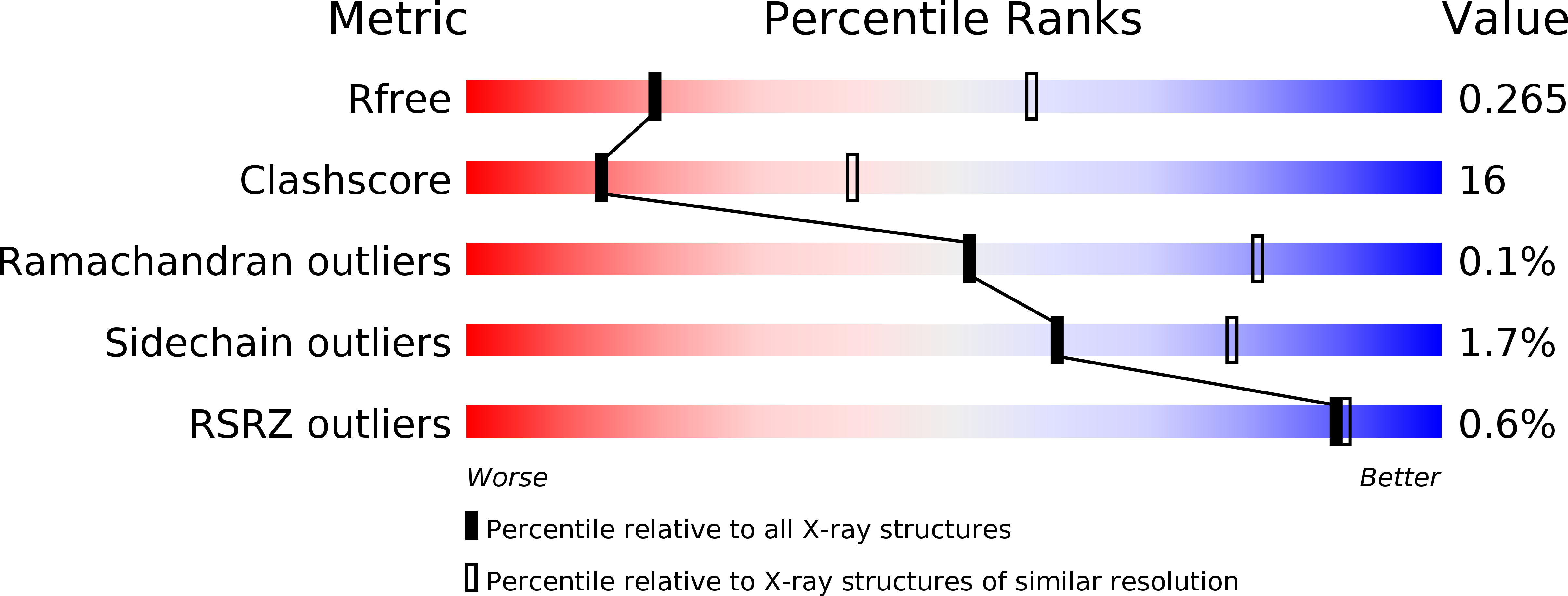
R-Value Free:
0.26
R-Value Work:
0.21
R-Value Observed:
0.21
Space Group:
P 1 21 1