3QHS image
Deposition Date 2011-01-26
Release Date 2011-05-18
Last Version Date 2023-11-01
Entry Detail
PDB ID:
3QHS
Title:
Crystal structure of full-length Hfq from Escherichia coli
Biological Source:
Source Organism:
Host Organism:
Method Details:
Experimental Method:
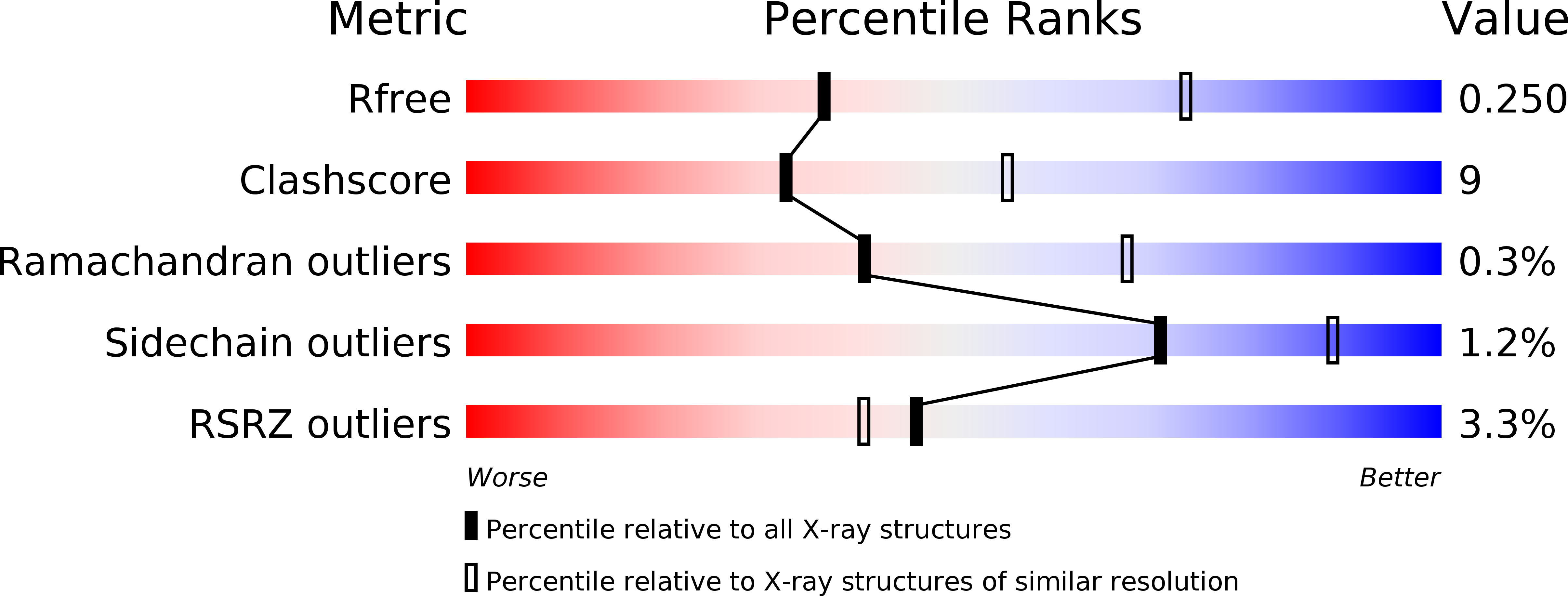
Resolution:
2.85 Å
R-Value Free:
0.24
R-Value Work:
0.20
R-Value Observed:
0.20
Space Group:
P 1
Macromolecular Entities
Structures with similar UniProt ID
Protein Blast
Polymer Type:polypeptide(L)
Molecule:Protein hfq
Gene (Uniprot):hfq
Chain IDs:A, B, C, D, E, F, G, H, I, J, K, L
Chain Length:102
Number of Molecules:12
Biological Source:Escherichia coli
Primary Citation
Structural analysis of full-length Hfq from Escherichia coli
Acta Crystallogr.,Sect.F 67 536 540 (2011)
PMID: 21543856 DOI: 10.1107/S174430911100786X

Abstact

The structure of full-length host factor Qβ (Hfq) from Escherichia coli obtained from a crystal belonging to space group P1, with unit-cell parameters a = 61.91, b = 62.15, c = 81.26 Å, α = 78.6, β = 86.2, γ = 59.9°, was solved by molecular replacement to a resolution of 2.85 Å and refined to R(work) and R(free) values of 20.7% and 25.0%, respectively. Hfq from E. coli has previously been crystallized and the structure has been solved for the N-terminal 72 amino acids, which cover ~65% of the full-length sequence. Here, the purification, crystallization and structural data of the full 102-amino-acid protein are presented. These data revealed that the presence of the C-terminus changes the crystal packing of E. coli Hfq. The crystal structure is discussed in the context of the recently published solution structure of Hfq from E. coli.

Legend

Protein

Chemical

Disease

Primary Citation of related structures