3QGL image
Deposition Date 2011-01-24
Release Date 2011-03-16
Last Version Date 2023-09-13
Entry Detail
PDB ID:
3QGL
Keywords:
Title:
Crystal Structure of PDZ domain of sorting nexin 27 (SNX27) in complex with the ESESKV peptide corresponding to the C-terminal tail of GIRK3
Biological Source:
Source Organism:
Host Organism:
Method Details:
Experimental Method:
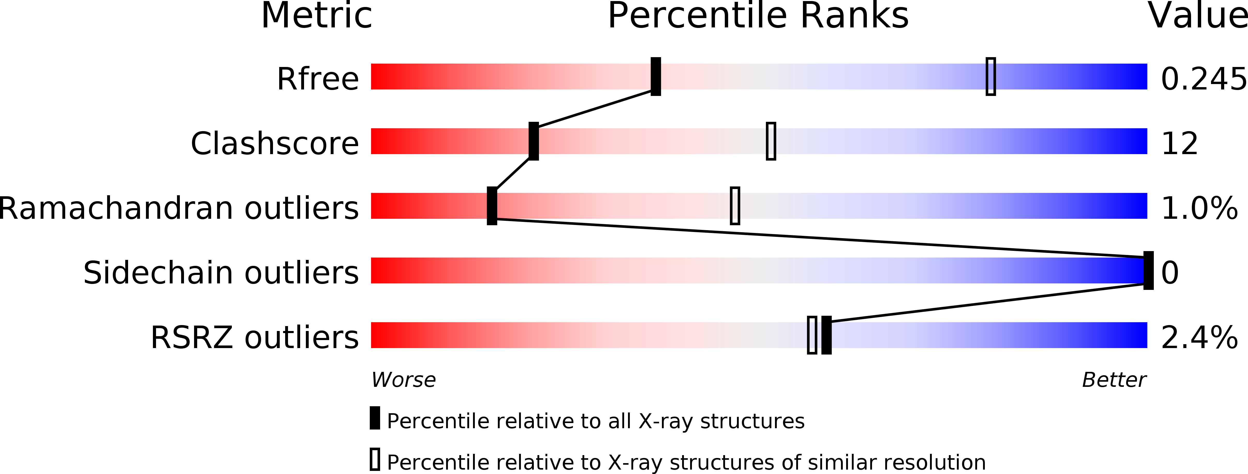
Resolution:
3.31 Å
R-Value Free:
0.25
R-Value Work:
0.23
R-Value Observed:
0.23
Space Group:
P 1 21 1
Macromolecular Entities
Polymer Type:polypeptide(L)
Molecule:Sorting nexin-27
Gene (Uniprot):Snx27
Chain IDs:A, C (auth: B), E (auth: C), G (auth: D), I (auth: E)
Chain Length:101
Number of Molecules:5
Biological Source:Rattus norvegicus
Polymer Type:polypeptide(L)
Molecule:G protein-activated inward rectifier potassium channel 3
Gene (Uniprot):Kcnj9
Chain IDs:B (auth: F), D (auth: G), F (auth: H), H (auth: I), J
Chain Length:6
Number of Molecules:5
Biological Source:Rattus norvegicus
Primary Citation
Mechanism underlying selective regulation of G protein-gated inwardly rectifying potassium channels by the psychostimulant-sensitive sorting nexin 27.
Proc.Natl.Acad.Sci.USA 108 5831 5836 (2011)
PMID: 21422294 DOI: 10.1073/pnas.1018645108

Abstact

G protein-gated inwardly rectifying potassium (GIRK) channels are important gatekeepers of neuronal excitability. The surface expression of neuronal GIRK channels is regulated by the psychostimulant-sensitive sorting nexin 27 (SNX27) protein through a class I (-X-Ser/Thr-X-Φ, where X is any residue and Φ is a hydrophobic amino acid) PDZ-binding interaction. The G protein-insensitive inward rectifier channel (IRK1) contains the same class I PDZ-binding motif but associates with a different synaptic PDZ protein, postsynaptic density protein 95 (PSD95). The mechanism by which SNX27 and PSD95 discriminate these channels was previously unclear. Using high-resolution structures coupled with biochemical and functional analyses, we identified key amino acids upstream of the channel's canonical PDZ-binding motif that associate electrostatically with a unique structural pocket in the SNX27-PDZ domain. Changing specific charged residues in the channel's carboxyl terminus or in the PDZ domain converts the selective association and functional regulation by SNX27. Elucidation of this unique interaction site between ion channels and PDZ-containing proteins could provide a therapeutic target for treating brain diseases.

Legend

Protein

Chemical

Disease

Primary Citation of related structures