
Deposition Date
2011-01-21
Release Date
2011-09-14
Last Version Date
2024-04-03
Entry Detail
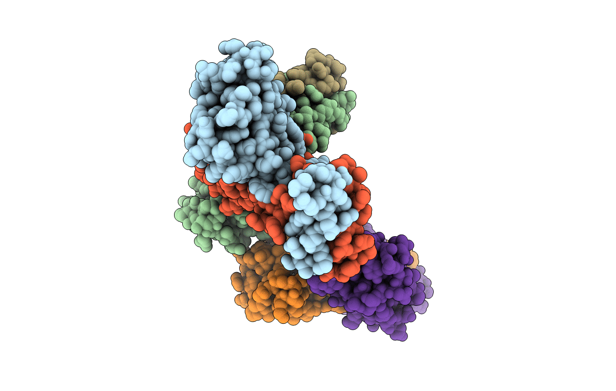
PDB ID:
3QF3
Keywords:
Title:
Crystal structure of EspR transcription factor from mycobacterium tuberculosis
Biological Source:
Source Organism(s):
Mycobacterium tuberculosis (Taxon ID: 1773)
Expression System(s):
Method Details:
Experimental Method:
Resolution:
2.41 Å
R-Value Free:
0.24
R-Value Work:
0.18
R-Value Observed:
0.18
Space Group:
P 1 21 1
