Deposition Date
2011-01-20
Release Date
2011-04-20
Last Version Date
2024-02-21
Entry Detail
PDB ID:
3QEB
Keywords:
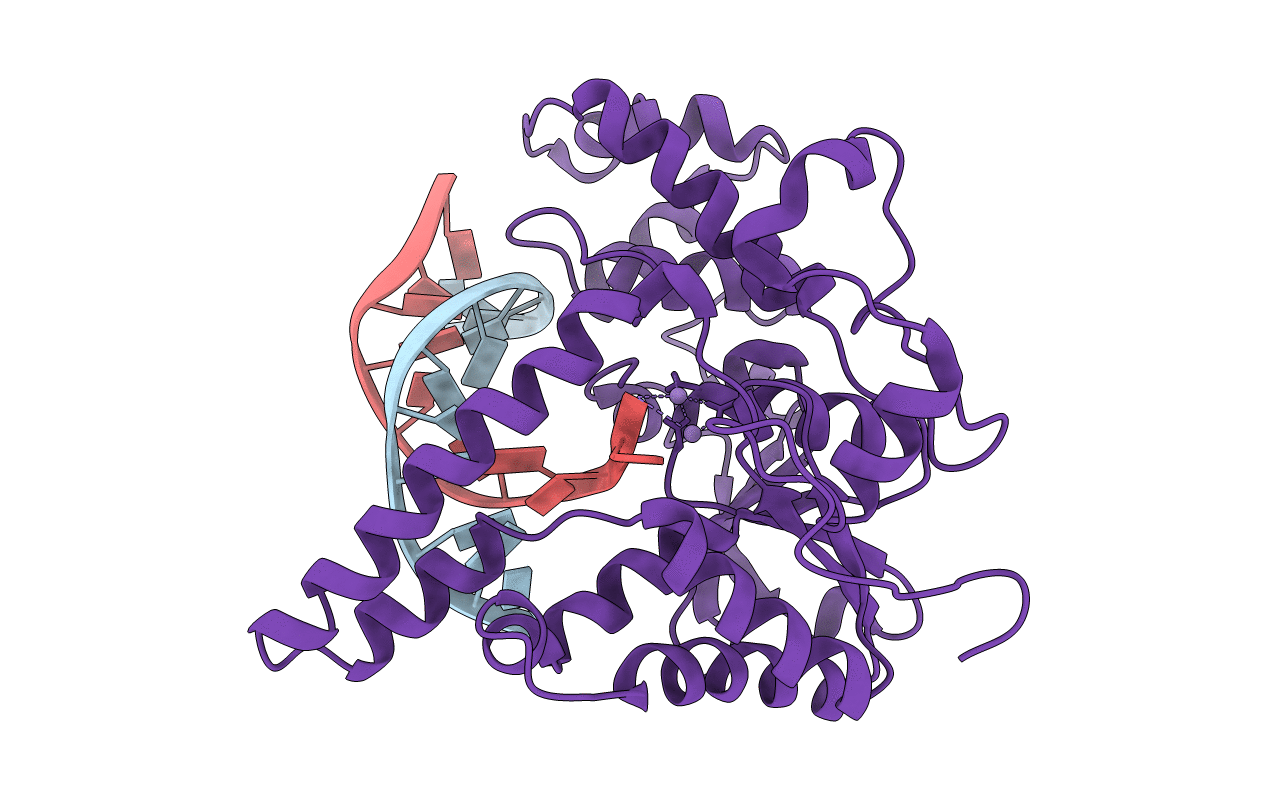
Title:
Crystal structure of human exonuclease 1 Exo1 (WT) in complex with DNA and Mn2+ (complex III)
Biological Source:
Source Organism(s):
Homo sapiens (Taxon ID: 9606)
Expression System(s):
Method Details:
Experimental Method:
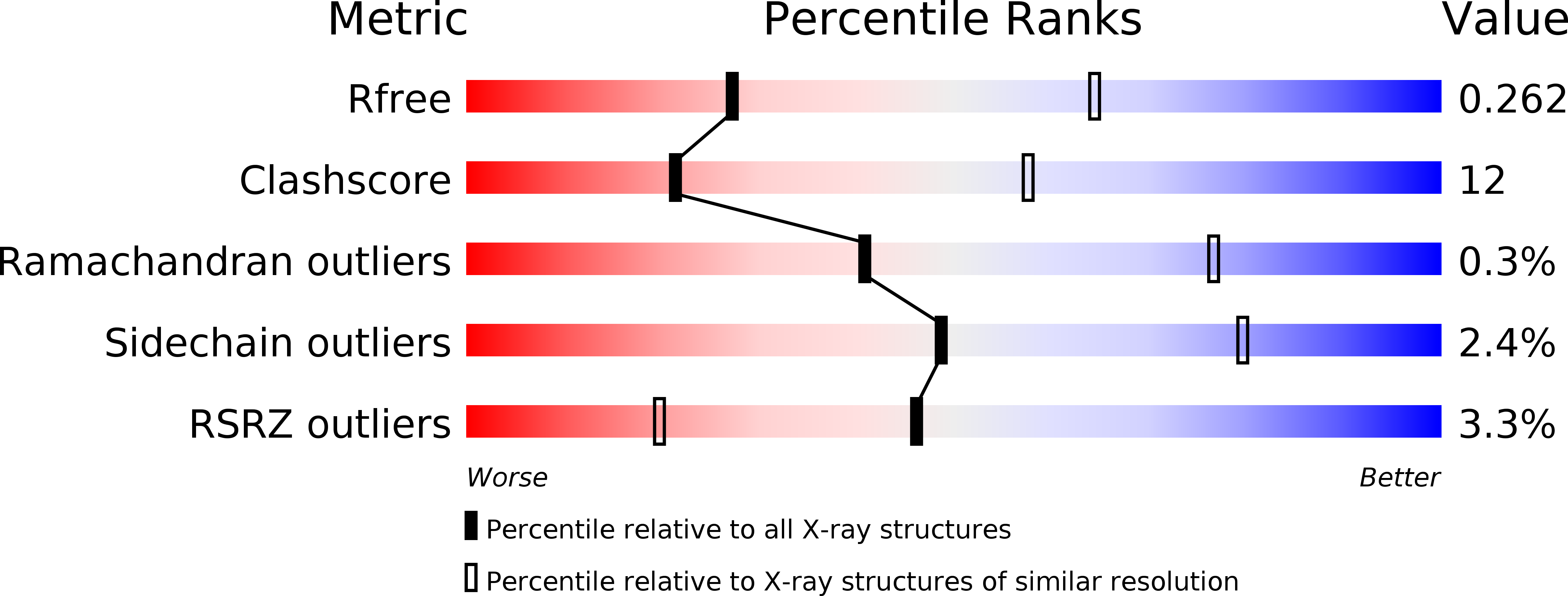
Resolution:
3.00 Å
R-Value Free:
0.26
R-Value Work:
0.22
R-Value Observed:
0.22
Space Group:
P 43 21 2