3QE5 image
Entry Detail
PDB ID:
3QE5
Keywords:
Title:
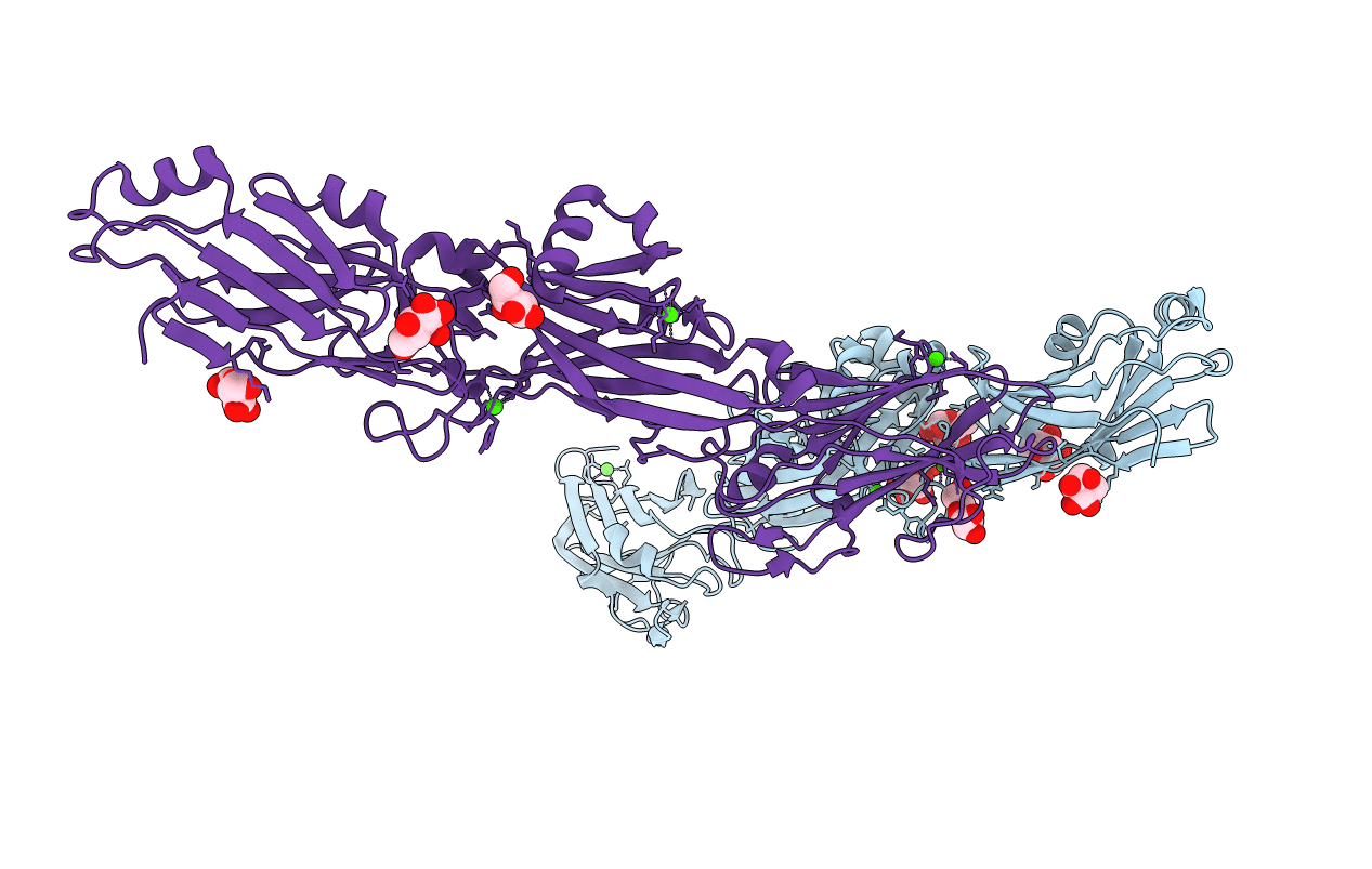
Complete structure of Streptococcus mutans Antigen I/II carboxy-terminus
Biological Source:
Source Organism:
Host Organism:
PDB Version:
Deposition Date:
2011-01-19
Release Date:
2011-04-20
Method Details:
Experimental Method:
Resolution:
2.50 Å
R-Value Free:
0.24
R-Value Work:
0.20
R-Value Observed:
0.20
Space Group:
I 2 2 2
Macromolecular Entities
Polymer Type:polypeptide(L)
Description:Major cell-surface adhesin PAc
Chain IDs:A, B
Chain Length:516
Number of Molecules:2
Biological Source:Streptococcus mutans
Primary Citation
Crystal Structure of the C-terminal Region of Streptococcus mutans Antigen I/II and Characterization of Salivary Agglutinin Adherence Domains.
J.Biol.Chem. 286 21657 21666 (2011)
PMID: 21505225 DOI: 10.1074/jbc.M111.231100

Abstact

The Streptococcus mutans antigen I/II (AgI/II) is a cell surface-localized protein that adheres to salivary components and extracellular matrix molecules. Here we report the 2.5 Å resolution crystal structure of the complete C-terminal region of AgI/II. The C-terminal region is comprised of three major domains: C(1), C(2), and C(3). Each domain adopts a DE-variant IgG fold, with two β-sheets whose A and F strands are linked through an intramolecular isopeptide bond. The adherence of the C-terminal AgI/II fragments to the putative tooth surface receptor salivary agglutinin (SAG), as monitored by surface plasmon resonance, indicated that the minimal region of binding was contained within the first and second DE-variant-IgG domains (C(1) and C(2)) of the C terminus. The minimal C-terminal region that could inhibit S. mutans adherence to SAG was also confirmed to be within the C(1) and C(2) domains. Competition experiments demonstrated that the C- and N-terminal regions of AgI/II adhere to distinct sites on SAG. A cleft formed at the intersection between these C(1) and C(2) domains bound glucose molecules from the cryo-protectant solution, revealing a putative binding site for its highly glycosylated receptor SAG. Finally, electron microscopy images confirmed the elongated structure of AgI/II and enabled building a composite tertiary model that encompasses its two distinct binding regions.

Legend

Protein

Chemical

Disease

Primary Citation of related structures