Planned Maintenance: Some services may turn out to be unavailable from 15th January, 2026 to 16th January, 2026. We apologize for the inconvenience!

3QDK image
Deposition Date 2011-01-18
Release Date 2011-02-09
Last Version Date 2024-02-21
Entry Detail
PDB ID:
3QDK
Keywords:
Title:
Structural insight on mechanism and diverse substrate selection strategy of ribulokinase
Method Details:
Experimental Method:
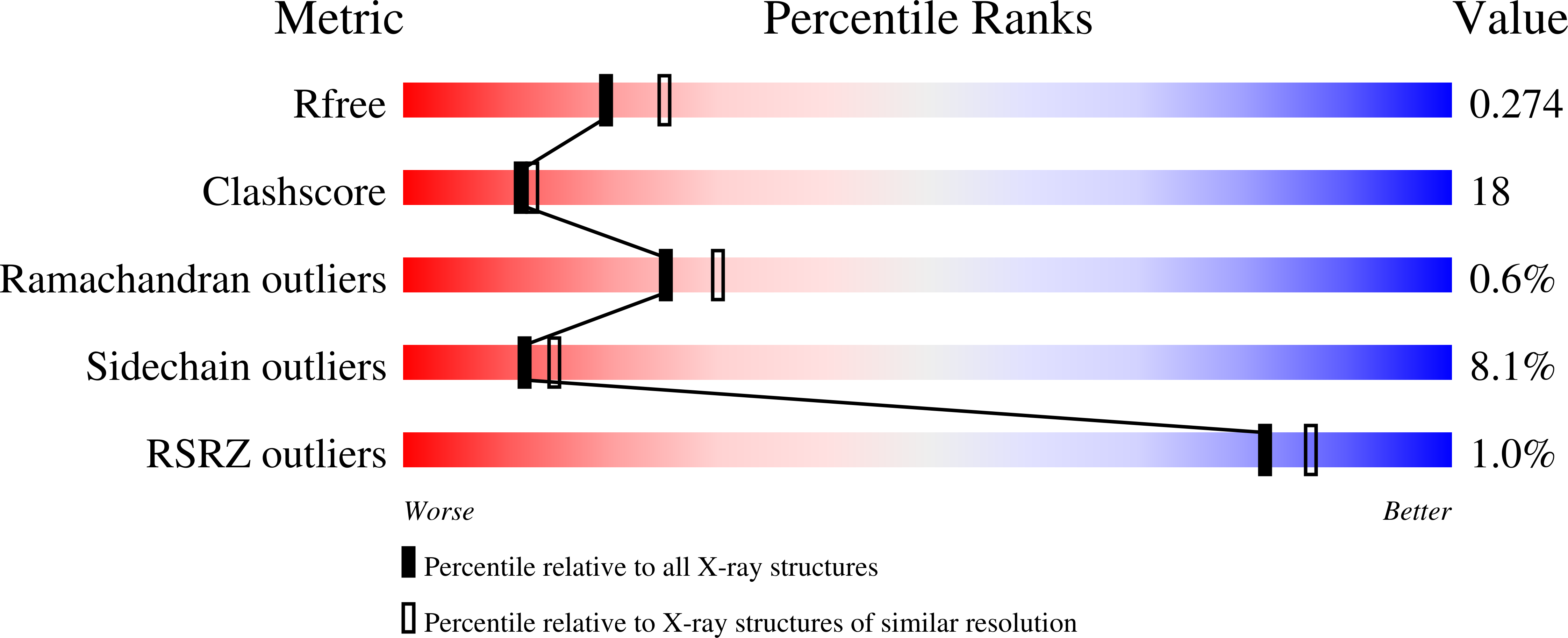
Resolution:
2.31 Å
R-Value Free:
0.27
R-Value Work:
0.22
R-Value Observed:
0.22
Space Group:
P 1 21 1
Macromolecular Entities
Structures with similar UniProt ID
Protein Blast
Polymer Type:polypeptide(L)
Molecule:Ribulokinase
Gene (Uniprot):araB
Chain IDs:A, B, C, D
Chain Length:572
Number of Molecules:4
Biological Source:Bacillus halodurans
Ligand Molecules
Primary Citation
Structural insight into mechanism and diverse substrate selection strategy of L-ribulokinase.
Proteins 80 261 268 (2012)
PMID: 22072612 DOI: 10.1002/prot.23202

Abstact

The araBAD operon encodes three different enzymes required for catabolism of L-arabinose, which is one of the most abundant monosaccharides in nature. L-ribulokinase, encoded by the araB gene, catalyzes conversion of L-ribulose to L-ribulose-5-phosphate, the second step in the catabolic pathway. Unlike other kinases, ribulokinase exhibits diversity in substrate selectivity and catalyzes phosphorylation of all four 2-ketopentose sugars with comparable k(cat) values. To understand ribulokinase recognition and phosphorylation of a diverse set of substrates, we have determined the X-ray structure of ribulokinase from Bacillus halodurans bound to L-ribulose and investigated its substrate and ATP co-factor binding properties. The polypeptide chain is folded into two domains, one small and the other large, with a deep cleft in between. By analogy with related sugar kinases, we identified (447)GGLPQK(452) as the ATP-binding motif within the smaller domain. L-ribulose binds in the cleft between the two domains via hydrogen bonds with the side chains of highly conserved Trp126, Lys208, Asp274, and Glu329 and the main chain nitrogen of Ala96. The interaction of L-ribulokinase with L-ribulose reveals versatile structural features that help explain recognition of various 2-ketopentose substrates and competitive inhibition by L-erythrulose. Comparison of our structure to that of the structures of other sugar kinases revealed conformational variations that suggest domain-domain closure movements are responsible for establishing the observed active site environment.

Legend

Protein

Chemical

Disease

Primary Citation of related structures
Feedback Form
Name
Email
Institute
Feedback