3QD7 image
Deposition Date 2011-01-18
Release Date 2011-06-22
Last Version Date 2024-03-20
Entry Detail
PDB ID:
3QD7
Keywords:
Title:
Crystal structure of YdaL, a stand-alone small MutS-related protein from Escherichia coli
Biological Source:
Source Organism(s):
Expression System(s):
Method Details:
Experimental Method:
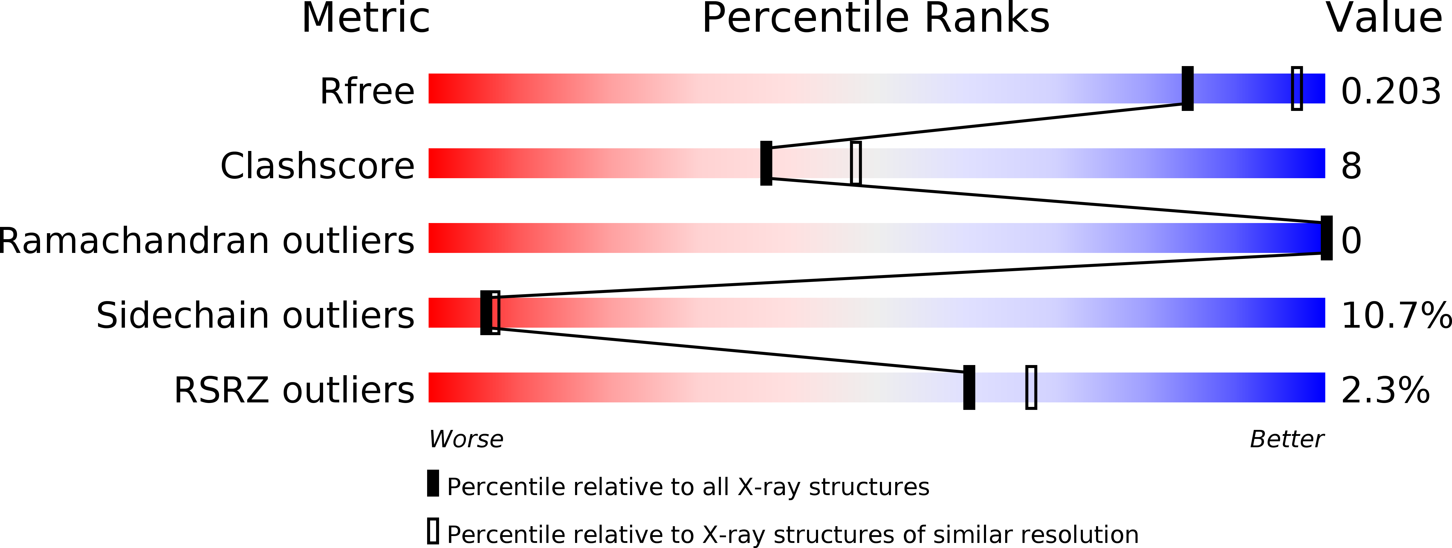
Resolution:
2.30 Å
R-Value Free:
0.24
R-Value Work:
0.22
R-Value Observed:
0.22
Space Group:
P 21 21 2
Macromolecular Entities
Structures with similar UniProt ID
Protein Blast
Polymer Type:polypeptide(L)
Molecule:Uncharacterized protein ydaL
Gene (Uniprot):smrA
Chain IDs:A (auth: X)
Chain Length:137
Number of Molecules:1
Biological Source:Escherichia coli
Primary Citation
Crystal structure of YdaL, a stand-alone small MutS-related protein from Escherichia coli.
J.Struct.Biol. 174 282 289 (2011)
PMID: 21276852 DOI: 10.1016/j.jsb.2011.01.008

Abstact

Sequence homologs of the small MutS-related (Smr) domain, the C-terminal endonuclease domain of MutS2, also exist as stand-alone proteins. In this study, we report the crystal structure of a proteolyzed fragment of YdaL (YdaL₃₉-₁₇₅), a stand-alone Smr protein from Escherichia coli. In this structure, residues 86-170 assemble into a classical Smr core domain and are embraced by an N-terminal extension (residues 40-85) with an α/β/α fold. Sequence alignment indicates that the N-terminal extension is conserved among a number of stand-alone Smr proteins, suggesting structural diversity among Smr domains. We also discovered that the DNA binding affinity and endonuclease activity of the truncated YdaL₃₉-₁₇₅ protein were slightly lower than those of full-length YdaL₁-₁₈₇, suggesting that residues 1-38 may be involved in DNA binding.

Legend

Protein

Chemical

Disease

Primary Citation of related structures
Feedback Form
Name
Email
Institute
Feedback