3QCA image
Deposition Date 2011-01-15
Release Date 2011-05-25
Last Version Date 2024-03-20
Entry Detail
PDB ID:
3QCA
Keywords:
Title:
Crystal Structure of FAF1 UBX Domain In Complex with p97/VCP N Domain Reveals The Conserved FcisP Touch-Turn Motif of UBX Domain Suffering Conformational Change
Biological Source:
Source Organism:
Homo sapiens (Taxon ID: 9606)
Host Organism:
Method Details:
Experimental Method:
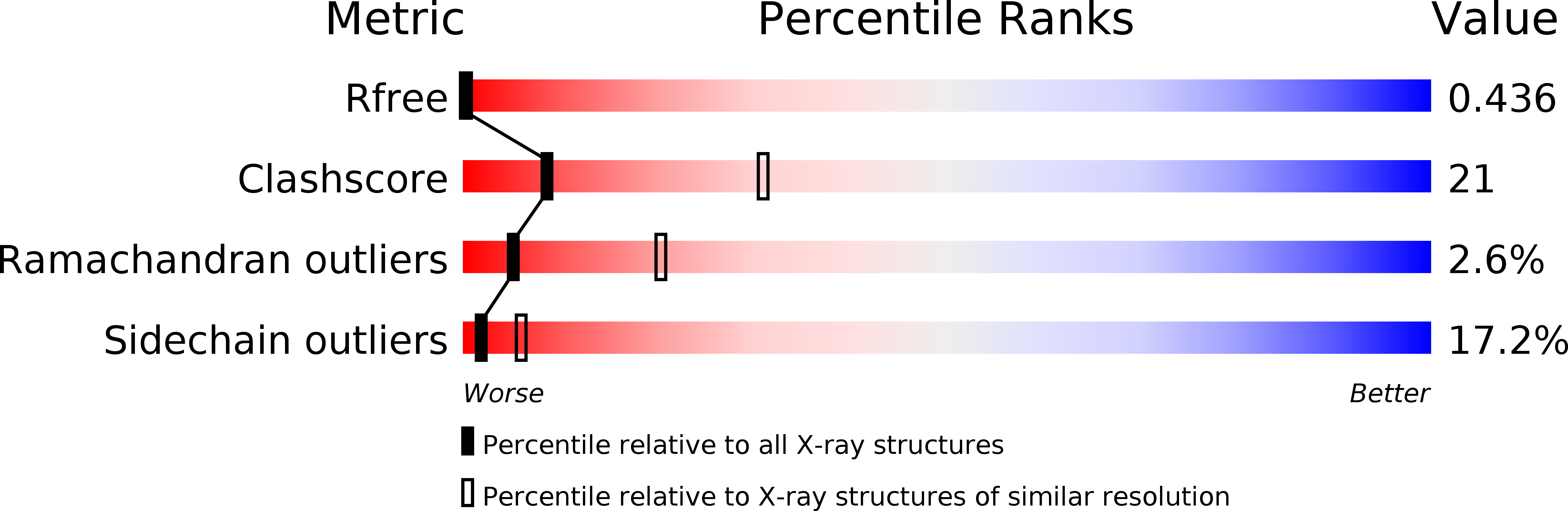
Resolution:
2.90 Å
R-Value Free:
0.29
R-Value Work:
0.24
R-Value Observed:
0.24
Space Group:
F 2 3
Macromolecular Entities
Structures with similar UniProt ID
Protein Blast
Polymer Type:polypeptide(L)
Molecule:FAS-associated factor 1
Gene (Uniprot):FAF1
Chain IDs:A, B, C, D
Chain Length:84
Number of Molecules:4
Biological Source:Homo sapiens
Primary Citation
Crystal structure of human FAF1 UBX domain reveals a novel FcisP touch-turn motif in p97/VCP-binding region
Biochem.Biophys.Res.Commun. 407 531 534 (2011)
PMID: 21414298 DOI: 10.1016/j.bbrc.2011.03.052

Abstact

UBX domain is a general p97/VCP-binding module found in an increasing number of proteins including FAF1, p47, SAKS1 and UBXD7. FAF1, a multi-functional tumor suppressor protein, binds to the N domain of p97/VCP through its C-terminal UBX domain and thereby inhibits the proteasomal protein degradation in which p97/VCP acts as a co-chaperone. Here we report the crystal structure of human FAF1 UBX domain at 2.9Å resolution. It reveals that the conserved FP sequence in the p97/VCP-binding region adopts a rarely observed cis-Pro touch-turn structure. We call it an FcisP touch-turn motif and suggest that it is the conserved structural element of the UBX domain. Four FAF1 UBX molecules in an asymmetric unit of the crystal show two different conformations of the FcisP touch-turn motif. The phenyl ring of F(619) in the motif stacks partly over cis-Pro(620) in one conformation, whereas it is swung out from cis-P(620), in the other conformation, and forms hydrophobic contacts with the residues of the neighboring molecule. In addition, the entire FcisP touch-turn motif is pulled out in the second conformation by about 2Å in comparison to the first conformation. Those conformational differences observed in the p97/VCP-binding motif caused by the interaction with neighboring molecules presumably represent the conformational change of the UBX domain on its binding to the N domain of p97/VCP.

Legend

Protein

Chemical

Disease

Primary Citation of related structures