Deposition Date
2011-01-10
Release Date
2011-11-30
Last Version Date
2024-10-09
Entry Detail
PDB ID:
3QA3
Keywords:
Title:
Crystal Structure of A-domain in complex with antibody
Biological Source:
Source Organism(s):
Homo sapiens (Taxon ID: 9606)
Mus musculus (Taxon ID: 10090)
Mus musculus (Taxon ID: 10090)
Expression System(s):
Method Details:
Experimental Method:
Resolution:
3.00 Å
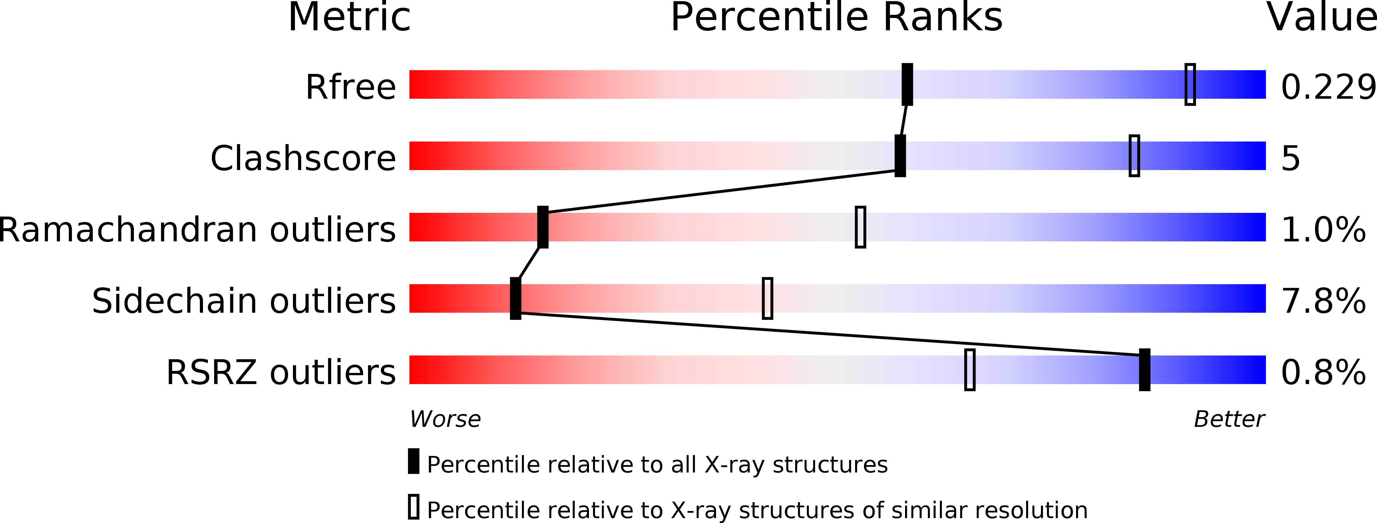
R-Value Free:
0.22
R-Value Work:
0.19
R-Value Observed:
0.19
Space Group:
P 21 21 21