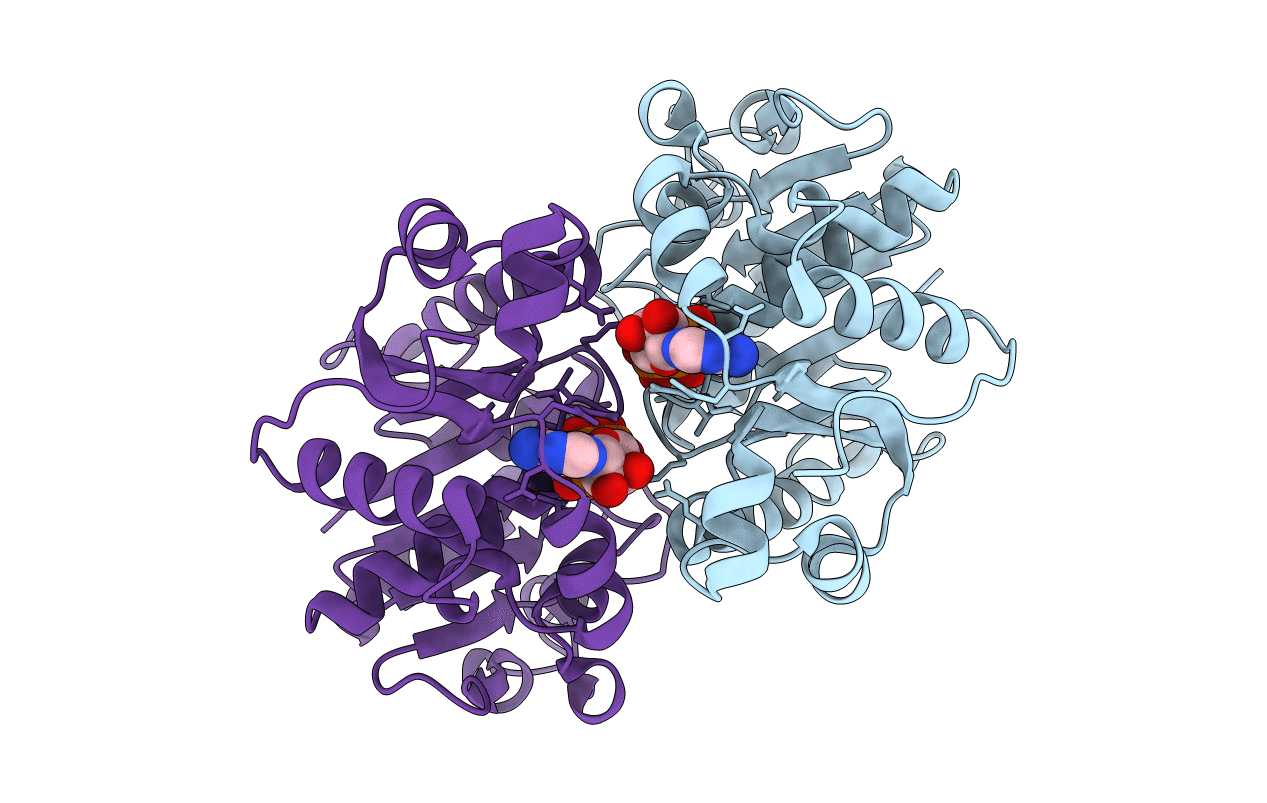
3Q9L image
Entry Detail
PDB ID:
3Q9L
Title:
The structure of the dimeric E.coli MinD-ATP complex
Biological Source:
Source Organism:
Host Organism:
PDB Version:
Deposition Date:
2011-01-08
Release Date:
2011-01-26
Method Details:
Experimental Method:
Resolution:
2.34 Å
R-Value Free:
0.30
R-Value Work:
0.26
R-Value Observed:
0.26
Space Group:
P 21 21 21
Macromolecular Entities
Polymer Type:polypeptide(L)
Description:Septum site-determining protein minD
Mutations:D40A
Chain IDs:A, B
Chain Length:260
Number of Molecules:2
Biological Source:Escherichia coli
Primary Citation
Determination of the structure of the MinD-ATP complex reveals the orientation of MinD on the membrane and the relative location of the binding sites for MinE and MinC.
Mol.Microbiol. 79 1515 1528 (2011)
PMID: 21231967 DOI: 10.1111/j.1365-2958.2010.07536.x

Abstact

The three Min proteins spatially regulate Z ring positioning in Escherichia coli and are dynamically associated with the membrane. MinD binds to vesicles in the presence of ATP and can recruit MinC or MinE. Biochemical and genetic evidence indicate the binding sites for these two proteins on MinD overlap. Here we solved the structure of a hydrolytic-deficient mutant of MinD truncated for the C-terminal amphipathic helix involved in binding to the membrane. The structure solved in the presence of ATP is a dimer and reveals the face of MinD abutting the membrane. Using a combination of random and extensive site-directed mutagenesis additional residues important for MinE and MinC binding were identified. The location of these residues on the MinD structure confirms that the binding sites overlap and reveals that the binding sites are at the dimer interface and exposed to the cytosol. The location of the binding sites at the dimer interface offers a simple explanation for the ATP dependence of MinC and MinE binding to MinD.

Legend

Protein

Chemical

Disease

Primary Citation of related structures