3Q86 image
Deposition Date 2011-01-06
Release Date 2011-07-27
Last Version Date 2023-11-01
Entry Detail
PDB ID:
3Q86
Keywords:
Title:
Crystal structure of Staphylococcus aureus nucleoside diphosphate kinase complexed with GTP
Biological Source:
Source Organism:
Host Organism:
Method Details:
Experimental Method:
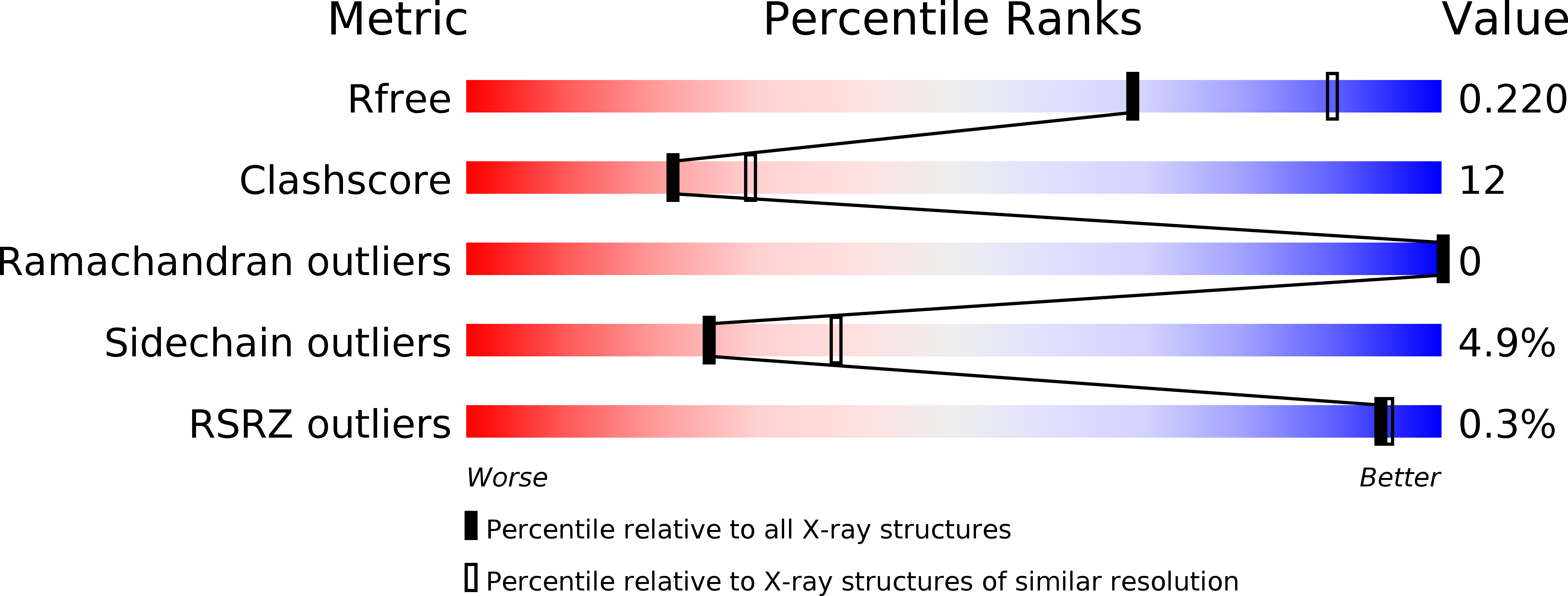
Resolution:
2.38 Å
R-Value Free:
0.22
R-Value Work:
0.16
R-Value Observed:
0.16
Space Group:
H 3
Macromolecular Entities
Structures with similar UniProt ID
Protein Blast
Polymer Type:polypeptide(L)
Molecule:Nucleoside diphosphate kinase
Gene (Uniprot):ndk
Chain IDs:A, B
Chain Length:157
Number of Molecules:2
Biological Source:Staphylococcus aureus subsp. aureus
Primary Citation
Conformational basis for substrate recognition and regulation of catalytic activity in Staphylococcus aureus nucleoside di-phosphate kinase.
Biochim.Biophys.Acta 1814 1349 1357 (2011)
PMID: 21745603 DOI: 10.1016/j.bbapap.2011.06.008

Abstact

Nucleoside diphosphate kinases (NDK) are characterized by high catalytic turnover rates and diverse substrate specificity. These features make this enzyme an effective activator of a pro-drug-an application that has been actively pursued for a variety of therapeutic strategies. The catalytic mechanism of this enzyme is governed by a conserved histidine that coordinates a magnesium ion at the active site. Despite substantial structural and biochemical information on NDK, the mechanistic feature of the phospho-transfer that leads to auto-phosphorylation remains unclear. While the role of the histidine residue is well documented, the other active site residues, in particular the conserved serine remains poorly characterized. Studies on some homologues suggest no role for the serine residue at the active site, while others suggest a crucial role for this serine in the regulation and quaternary association of this enzyme in some species. Here we report the biochemical features of the Staphylococcus aureus NDK and the mutant enzymes. We also describe the crystal structures of the apo-NDK, as a transition state mimic with vanadate and in complex with different nucleotide substrates. These structures formed the basis for molecular dynamics simulations to understand the broad substrate specificity of this enzyme and the role of active site residues in the phospho-transfer mechanism and oligomerization. Put together, these data suggest that concerted changes in the conformation of specific residues facilitate the stabilization of nucleotide complexes thereby enabling the steps involved in the ping-pong reaction mechanism without large changes to the overall structure of this enzyme.

Legend

Protein

Chemical

Disease

Primary Citation of related structures