3Q7Q image
Deposition Date 2011-01-05
Release Date 2011-09-21
Last Version Date 2023-09-13
Entry Detail
PDB ID:
3Q7Q
Title:
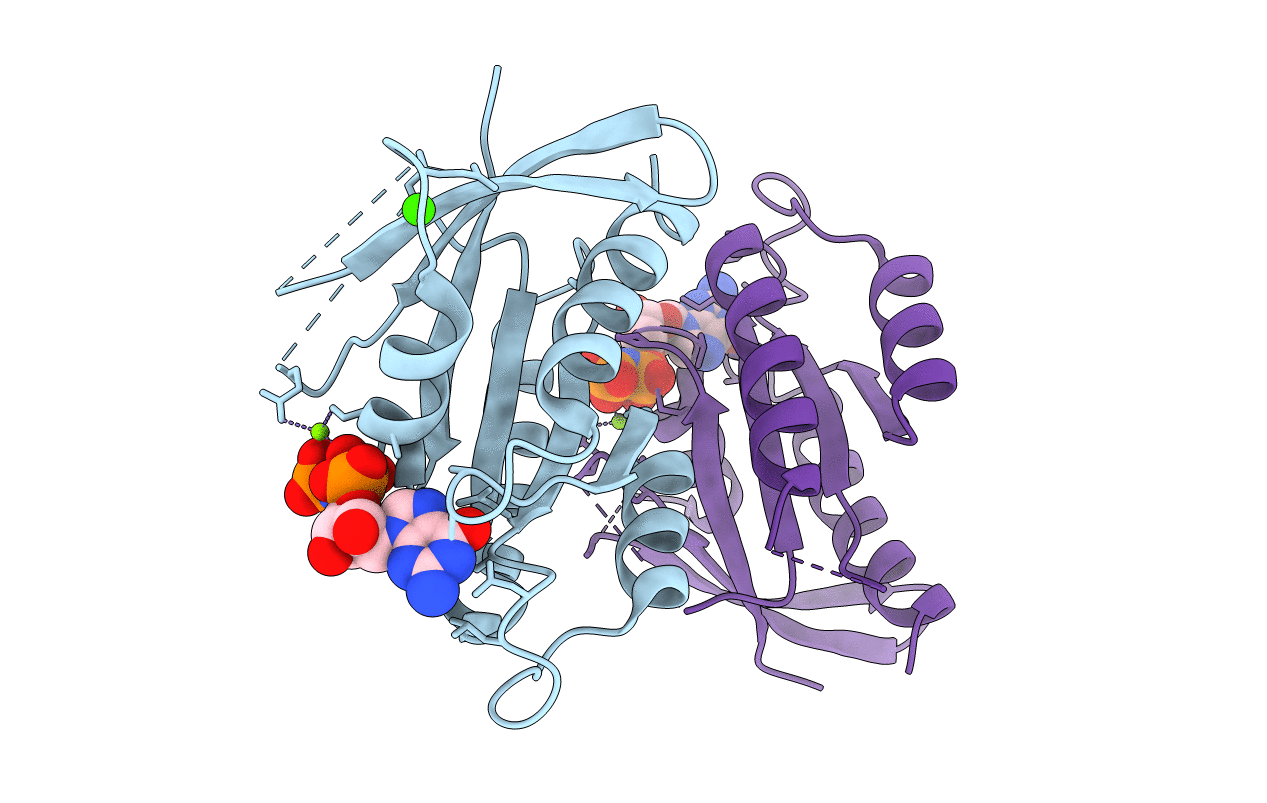
Crystal Structure of Rad G-domain Q148A-GTP Analog Complex
Biological Source:
Source Organism:
Homo sapiens (Taxon ID: 9606)
Host Organism:
Method Details:
Experimental Method:
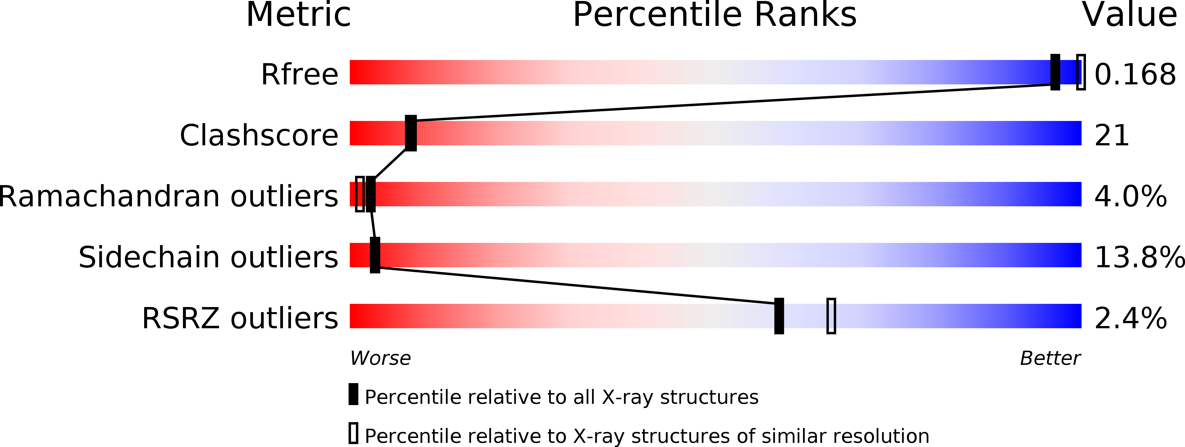
Resolution:
2.30 Å
R-Value Free:
0.20
R-Value Work:
0.16
R-Value Observed:
0.16
Space Group:
P 31
Macromolecular Entities
Polymer Type:polypeptide(L)
Molecule:GTP-binding protein RAD
Gene (Uniprot):RRAD
Mutations:Q148A
Chain IDs:A, B
Chain Length:166
Number of Molecules:2
Biological Source:Homo sapiens
Primary Citation
RGK Family G-Domain:GTP Analog Complex Structures and Nucleotide-Binding Properties.
J.Mol.Biol. 413 372 389 (2011)
PMID: 21903096 DOI: 10.1016/j.jmb.2011.08.017

Abstact

The RGK family of small G-proteins, including Rad, Gem, Rem1, and Rem2, is inducibly expressed in various mammalian tissues and interacts with voltage-dependent calcium channels and Rho kinase. Many questions remain regarding their physiological roles and molecular mechanism. Previous crystallographic studies reported RGK G-domain:guanosine di-phosphate structures. To test whether RGK proteins undergo a nucleotide-induced conformational change, we determined the crystallographic structures of Rad:GppNHp and Rem2:GppNHp to 1.7 and 1.8 Å resolutions, respectively. Also, we characterized the nucleotide-binding properties and conformations for Gem, Rad, and several structure-based mutants using fluorescence spectroscopy. The results suggest that RGK G-proteins may not behave as Ras-like canonical nucleotide-induced molecular switches. Further, the RGK proteins have differing structures and nucleotide-binding properties, which may have implications for their varied action on effectors.

Legend

Protein

Chemical

Disease

Primary Citation of related structures