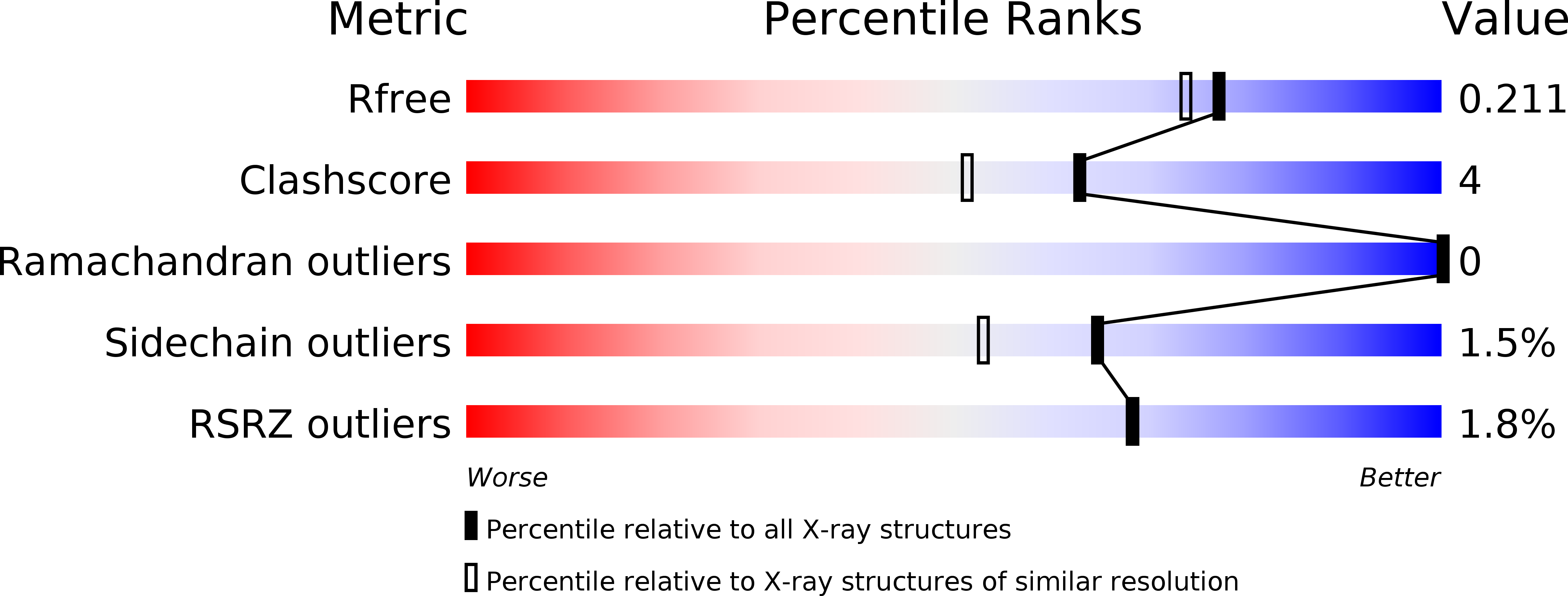
3Q76 image
Deposition Date 2011-01-04
Release Date 2011-05-11
Last Version Date 2024-10-30
Entry Detail
PDB ID:
3Q76
Keywords:
Title:
Structure of human neutrophil elastase (uncomplexed)
Biological Source:
Source Organism:
Homo sapiens (Taxon ID: 9606)
Method Details:
Experimental Method:
Resolution:
1.86 Å
R-Value Free:
0.21
R-Value Work:
0.17
R-Value Observed:
0.18
Space Group:
P 41 21 2
Macromolecular Entities
Structures with similar UniProt ID
Protein Blast
Polymer Type:polypeptide(L)
Molecule:Neutrophil elastase
Gene (Uniprot):ELANE
Chain IDs:A, B
Chain Length:218
Number of Molecules:2
Biological Source:Homo sapiens
Modified Residue
Compound ID Chain ID Parent Comp ID Details 2D Image
ASN A ASN GLYCOSYLATION SITE
Primary Citation
Unexpected active-site flexibility in the structure of human neutrophil elastase in complex with a new dihydropyrimidone inhibitor.
J.Mol.Biol. 409 681 691 (2011)
PMID: 21549129 DOI: 10.1016/j.jmb.2011.04.047

Abstact

Human neutrophil elastase (HNE), a trypsin-type serine protease, is of pivotal importance in the onset and progression of chronic obstructive pulmonary disease (COPD). COPD encompasses a group of slowly progressive respiratory disorders and is a major medical problem and the fifth leading cause of death worldwide. HNE is a major target for the development of compounds that inhibit the progression of long-term lung function decline in COPD patients. Here, we present the three-dimensional structure of a potent dihydropyrimidone inhibitor (DHPI) non-covalently bound to HNE at a resolution of 2.0 Å. The inhibitor binds to the active site in a unique orientation addressing S1 and S2 subsites of the protease. To facilitate further analysis of this binding mode, we determined the structure of the uncomplexed enzyme at a resolution of 1.86 Å. Detailed comparisons of the HNE:DHPI complex with the uncomplexed HNE structure and published structures of other elastase:inhibitor complexes revealed that binding of DHPI leads to large conformational changes in residues located in the S2 subsite. The rearrangement of residues Asp95-Leu99B creates a deep, well-defined cavity, which is filled by the P2 moiety of the inhibitor molecule to almost perfect shape complementarity. The shape of the S2 subsite in complex with DHPI clearly differs from all other observed HNE structures. The observed structural flexibility of the S2 subsite is a key feature for the understanding of the binding mode of DHPIs in general and the development of new HNE selective inhibitors.

Legend

Protein

Chemical

Disease

Primary Citation of related structures