Deposition Date
2010-12-28
Release Date
2011-01-19
Last Version Date
2024-11-20
Entry Detail
PDB ID:
3Q5D
Keywords:
Title:
crystal structure of human Atlastin-1 (residues 1-447) bound to GDP, crystal form 1
Biological Source:
Source Organism(s):
Homo sapiens (Taxon ID: 9606)
Expression System(s):
Method Details:
Experimental Method:
Resolution:
2.70 Å
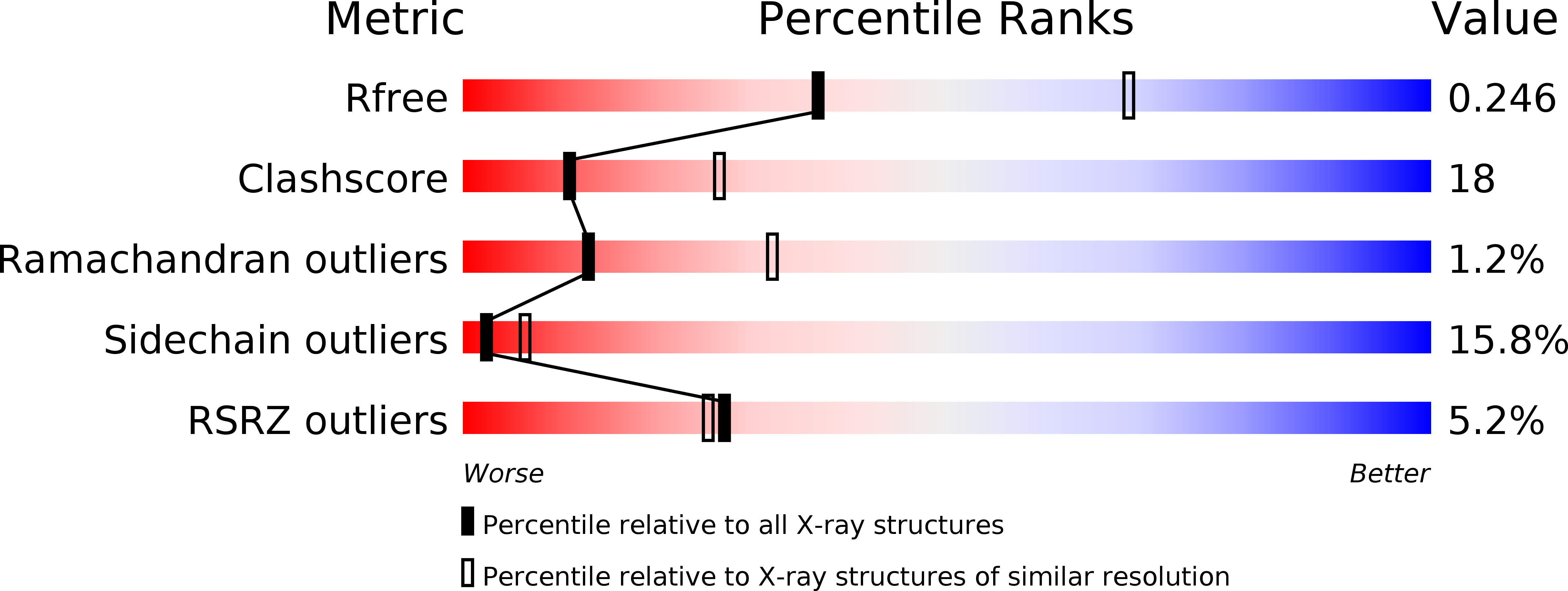
R-Value Free:
0.24
R-Value Work:
0.18
R-Value Observed:
0.19
Space Group:
P 65 2 2