
Deposition Date
2010-12-22
Release Date
2011-09-07
Last Version Date
2023-12-06
Entry Detail
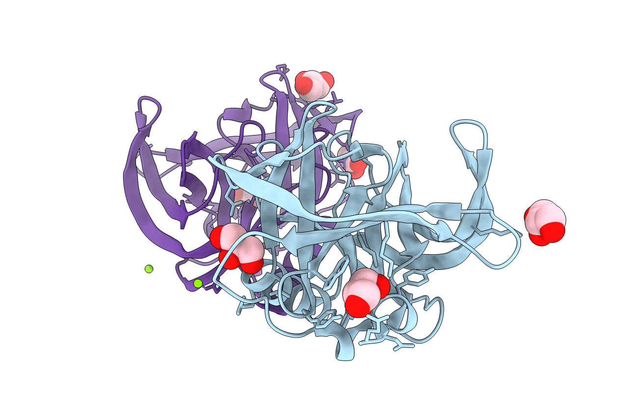
PDB ID:
3Q3X
Keywords:
Title:
Crystal structure of the main protease (3C) from human enterovirus B EV93
Biological Source:
Source Organism(s):
Human enterovirus B (Taxon ID: 138949)
Expression System(s):
Method Details:
Experimental Method:
Resolution:
1.90 Å
R-Value Free:
0.21
R-Value Work:
0.14
R-Value Observed:
0.15
Space Group:
P 1 21 1
