3Q2U image
Deposition Date 2010-12-20
Release Date 2011-10-05
Last Version Date 2024-10-16
Entry Detail
PDB ID:
3Q2U
Title:
Structure of Human Glioma Pathogenesis-related Protein 1 Reveals Unique loops and surface motifs.
Biological Source:
Source Organism:
Homo sapiens (Taxon ID: 9606)
Host Organism:
Method Details:
Experimental Method:
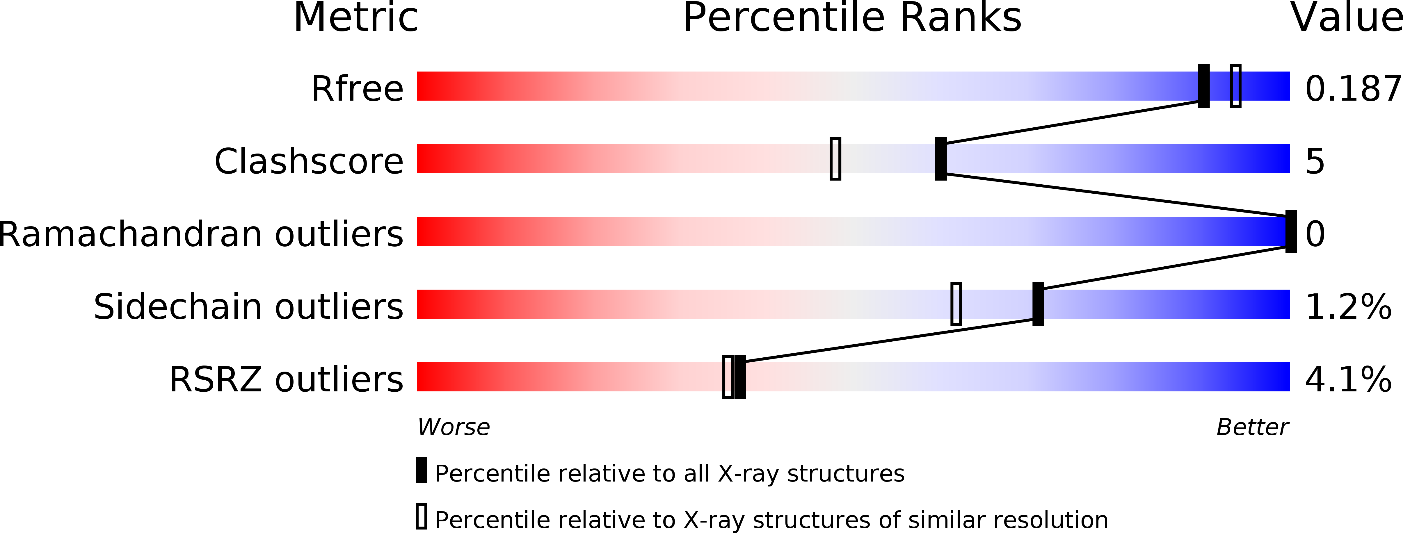
Resolution:
1.85 Å
R-Value Free:
0.18
R-Value Work:
0.13
R-Value Observed:
0.13
Space Group:
P 21 21 2
Macromolecular Entities
Polymer Type:polypeptide(L)
Molecule:Glioma pathogenesis-related protein 1
Gene (Uniprot):GLIPR1
Chain IDs:A
Chain Length:205
Number of Molecules:1
Biological Source:Homo sapiens
Ligand Molecules
Primary Citation
Structural studies of human glioma pathogenesis-related protein 1.
Acta Crystallogr.,Sect.D 67 847 855 (2011)
PMID: 21931216 DOI: 10.1107/S0907444911028198

Abstact

Human glioma pathogenesis-related protein 1 (GLIPR1) is a membrane protein that is highly upregulated in brain cancers but is barely detectable in normal brain tissue. GLIPR1 is composed of a signal peptide that directs its secretion, a conserved cysteine-rich CAP (cysteine-rich secretory proteins, antigen 5 and pathogenesis-related 1 proteins) domain and a transmembrane domain. GLIPR1 is currently being investigated as a candidate for prostate cancer gene therapy and for glioblastoma targeted therapy. Crystal structures of a truncated soluble domain of the human GLIPR1 protein (sGLIPR1) solved by molecular replacement using a truncated polyalanine search model of the CAP domain of stecrisp, a snake-venom cysteine-rich secretory protein (CRISP), are presented. The correct molecular-replacement solution could only be obtained by removing all loops from the search model. The native structure was refined to 1.85 Å resolution and that of a Zn2+ complex was refined to 2.2 Å resolution. The latter structure revealed that the putative binding cavity coordinates Zn2+ similarly to snake-venom CRISPs, which are involved in Zn2+-dependent mechanisms of inflammatory modulation. Both sGLIPR1 structures have extensive flexible loop/turn regions and unique charge distributions that were not observed in any of the previously reported CAP protein structures. A model is also proposed for the structure of full-length membrane-bound GLIPR1.

Legend

Protein

Chemical

Disease

Primary Citation of related structures