
Deposition Date
2010-12-15
Release Date
2011-02-23
Last Version Date
2024-02-21
Entry Detail
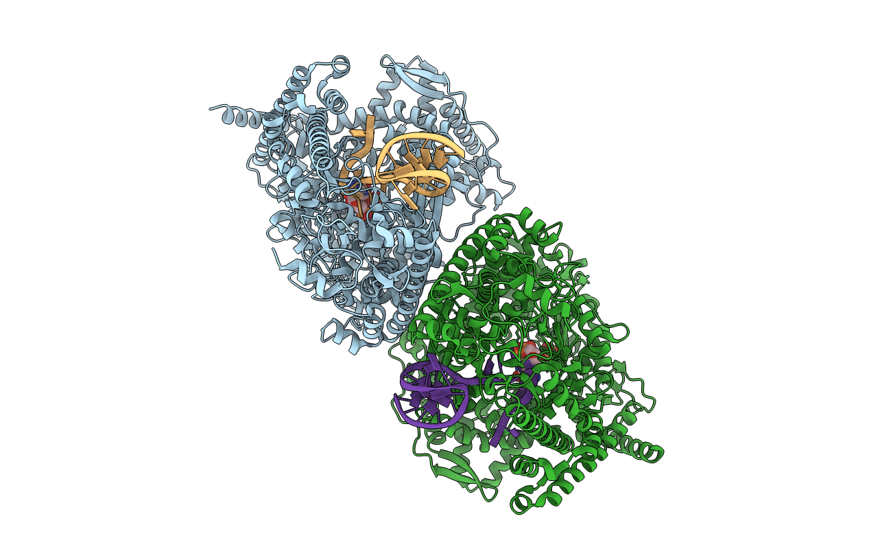
PDB ID:
3Q0A
Keywords:
Title:
X-ray crystal structure of the transcription initiation complex of the N4 mini-vRNAP with P2 promoter: Mismatch complex
Biological Source:
Source Organism(s):
Enterobacteria phage N4 (Taxon ID: 10752)
Expression System(s):
Method Details:
Experimental Method:
Resolution:
2.69 Å
R-Value Free:
0.27
R-Value Work:
0.21
R-Value Observed:
0.21
Space Group:
P 21 21 21
