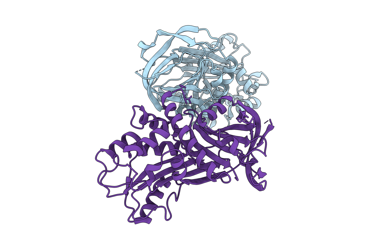
3Q03 image
Entry Detail
PDB ID:
3Q03
Title:
Crystal structure of plasminogen activator inhibitor-1 in a metastable active conformation.
Biological Source:
Source Organism:
Host Organism:
PDB Version:
Deposition Date:
2010-12-15
Release Date:
2011-06-22
Method Details:
Experimental Method:
Resolution:
2.64 Å
R-Value Free:
0.22
R-Value Work:
0.17
R-Value Observed:
0.17
Space Group:
P 1 21 1
Macromolecular Entities
Polymer Type:polypeptide(L)
Description:Plasminogen activator inhibitor 1
Mutations:W175F
Chain IDs:A, B
Chain Length:379
Number of Molecules:2
Biological Source:Homo sapiens
Primary Citation
Crystal structure of plasminogen activator inhibitor-1 in an active conformation with normal thermodynamic stability.
J.Biol.Chem. 286 29709 29717 (2011)
PMID: 21697084 DOI: 10.1074/jbc.M111.236554

Abstact

The serpin plasminogen activator inhibitor-1 (PAI-1) is a crucial regulator in fibrinolysis and tissue remodeling. PAI-1 has been associated with several pathological conditions and is a validated prognostic marker in human cancers. However, structural information about the native inhibitory form of PAI-1 has been elusive because of its inherent conformational instability and rapid conversion to a latent, inactive structure. Here we report the crystal structure of PAI-1 W175F at 2.3 Å resolution as the first model of the metastable native molecule. Structural comparison with a quadruple mutant (14-1B) previously used as representative of the active state uncovered key differences. The most striking differences occur near the region that houses three of the four mutations in the 14-1B PAI-1 structure. Prominent changes are localized within a loop connecting β-strand 3A with the F helix, in which a previously observed 3(10)-helix is absent in the new structure. Notably these structural changes are found near the binding site for the cofactor vitronectin. Because vitronectin is the only known physiological regulator of PAI-1 that slows down the latency conversion, the structure of this region is important. Furthermore, the previously identified chloride-binding site close to the F-helix is absent from the present structure and likely to be artifactual, because of its dependence on the 14-1B mutations. Instead we found a different chlorine-binding site that is likely to be present in wild type PAI-1 and that more satisfactorily accounts for the chlorine stabilizing effect on PAI-1.

Legend

Protein

Chemical

Disease

Primary Citation of related structures