3PYW image
Deposition Date 2010-12-13
Release Date 2011-04-27
Last Version Date 2024-02-21
Entry Detail
PDB ID:
3PYW
Title:
The structure of the SLH domain from B. anthracis surface array protein at 1.8A
Biological Source:
Source Organism:
Host Organism:
Method Details:
Experimental Method:
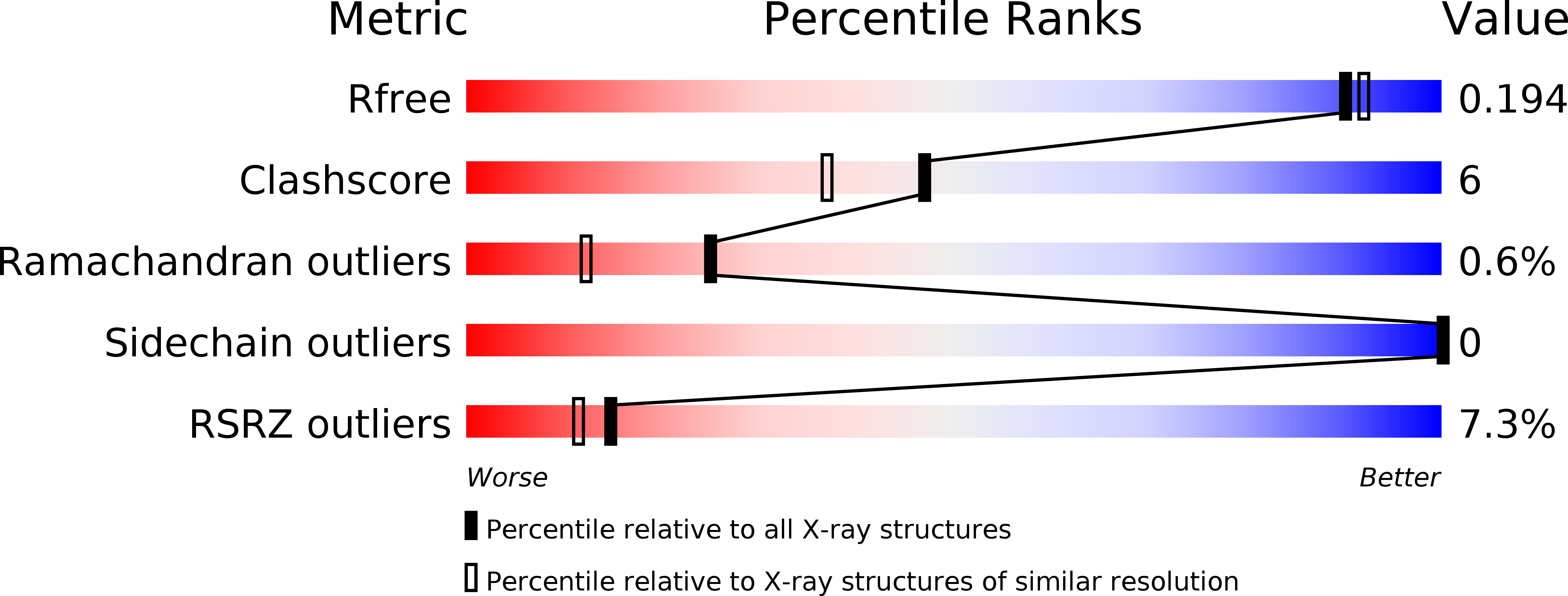
Resolution:
1.80 Å
R-Value Free:
0.18
R-Value Work:
0.16
R-Value Observed:
0.16
Space Group:
P 41 21 2
Macromolecular Entities
Structures with similar UniProt ID
Protein Blast
Polymer Type:polypeptide(L)
Molecule:S-layer protein sap
Gene (Uniprot):sap
Chain IDs:A
Chain Length:203
Number of Molecules:1
Biological Source:Bacillus anthracis
Ligand Molecules
Primary Citation
Structure of Surface Layer Homology (SLH) Domains from Bacillus anthracis Surface Array Protein.
J.Biol.Chem. 286 26042 26049 (2011)
PMID: 21572039 DOI: 10.1074/jbc.M111.248070

Abstact

Surface (S)-layers, para-crystalline arrays of protein, are deposited in the envelope of most bacterial species. These surface organelles are retained in the bacterial envelope through the non-covalent association of proteins with cell wall carbohydrates. Bacillus anthracis, a Gram-positive pathogen, produces S-layers of the protein Sap, which uses three consecutive repeats of the surface-layer homology (SLH) domain to engage secondary cell wall polysaccharides (SCWP). Using x-ray crystallography, we reveal here the structure of these SLH domains, which assume the shape of a three-prong spindle. Each SLH domain contributes to a three-helical bundle at the spindle base, whereas another α-helix and its connecting loops generate the three prongs. The inter-prong grooves contain conserved cationic and anionic residues, which are necessary for SLH domains to bind the B. anthracis SCWP. Modeling experiments suggest that the SLH domains of other S-layer proteins also fold into three-prong spindles and capture bacterial envelope carbohydrates by a similar mechanism.

Legend

Protein

Chemical

Disease

Primary Citation of related structures