3PY7 image
Deposition Date 2010-12-12
Release Date 2011-12-14
Last Version Date 2024-02-21
Entry Detail
PDB ID:
3PY7
Keywords:
Title:
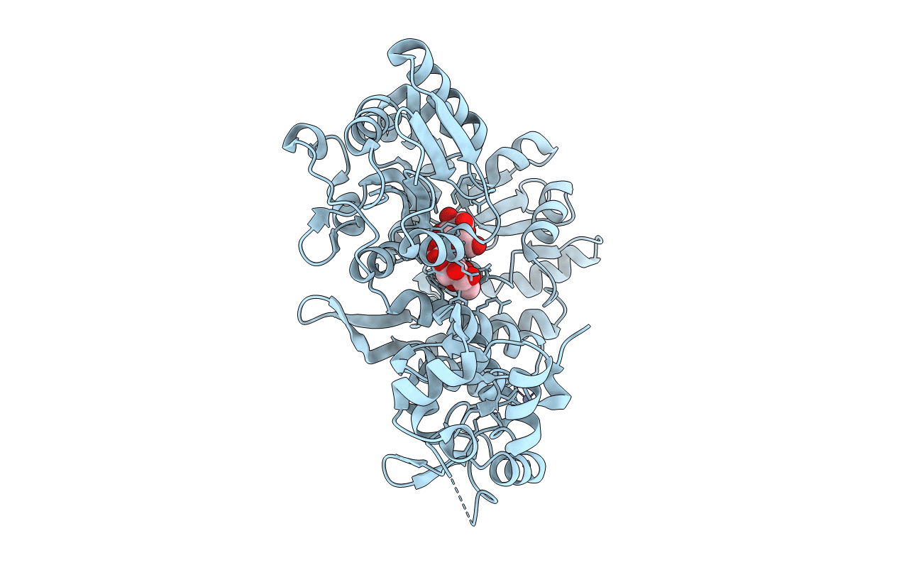
Crystal structure of full-length Bovine Papillomavirus oncoprotein E6 in complex with LD1 motif of paxillin at 2.3A resolution
Biological Source:
Source Organism:
Host Organism:
Method Details:
Experimental Method:
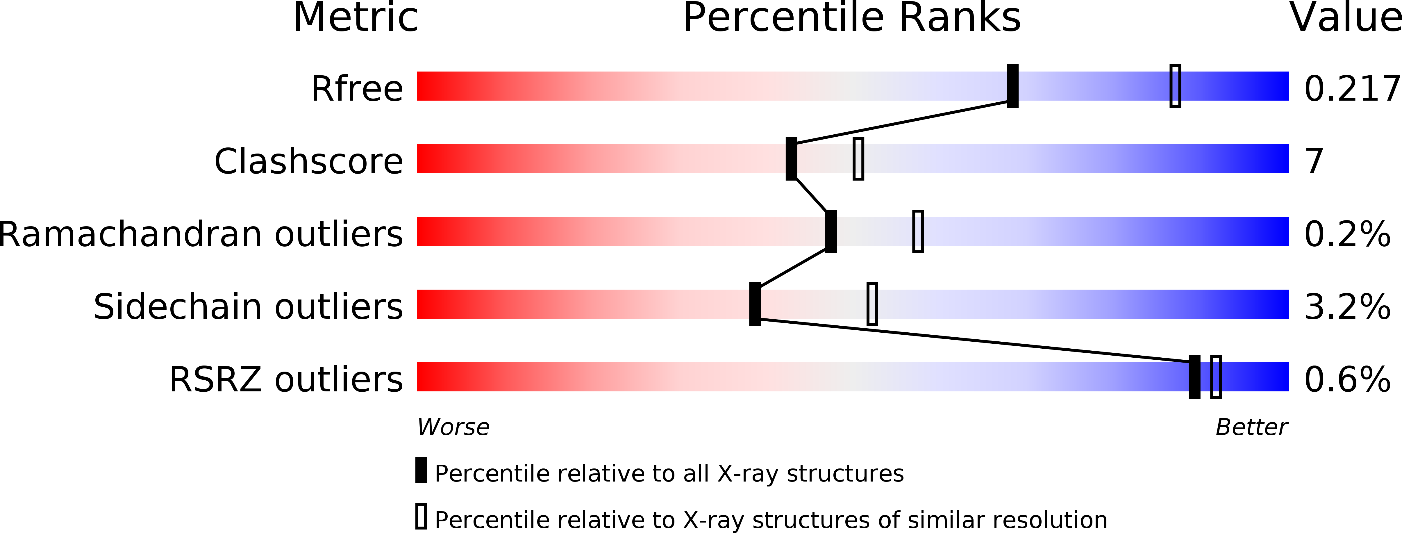
Resolution:
2.29 Å
R-Value Free:
0.22
R-Value Work:
0.18
R-Value Observed:
0.18
Space Group:
C 2 2 21
Macromolecular Entities
Polymer Type:polypeptide(L)
Molecule:maltose-binding periplasmic protein,paxillin LD1,protein E6 chimera
Gene (Uniprot):E6, malE
Mutations:D108A,K109A,K265A,E385A,K388A,D389A (maltose-binding periplasmic protein)
Chain IDs:A
Chain Length:523
Number of Molecules:1
Biological Source:Escherichia coli, bovine papillomavirus type 1, Homo sapiens
Ligand Molecules
Peptide-like Molecules
PRD_900009
Primary Citation
Structural basis for hijacking of cellular LxxLL motifs by papillomavirus E6 oncoproteins.
Science 339 694 698 (2013)
PMID: 23393263 DOI: 10.1126/science.1229934

Abstact

E6 viral oncoproteins are key players in epithelial tumors induced by papillomaviruses in vertebrates, including cervical cancer in humans. E6 proteins target many host proteins by specifically interacting with acidic LxxLL motifs. We solved the crystal structures of bovine (BPV1) and human (HPV16) papillomavirus E6 proteins bound to LxxLL peptides from the focal adhesion protein paxillin and the ubiquitin ligase E6AP, respectively. In both E6 proteins, two zinc domains and a linker helix form a basic-hydrophobic pocket, which captures helical LxxLL motifs in a way compatible with other interaction modes. Mutational inactivation of the LxxLL binding pocket disrupts the oncogenic activities of both E6 proteins. This work reveals the structural basis of both the multifunctionality and the oncogenicity of E6 proteins.

Legend

Protein

Chemical

Disease

Primary Citation of related structures