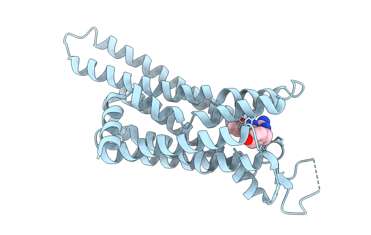
3PWH image
Entry Detail
PDB ID:
3PWH
Title:
Thermostabilised Adenosine A2A Receptor
Biological Source:
Source Organism:
Host Organism:
PDB Version:
Deposition Date:
2010-12-08
Release Date:
2011-09-07
Method Details:
Experimental Method:
Resolution:
3.30 Å
R-Value Free:
0.31
R-Value Work:
0.27
R-Value Observed:
0.27
Space Group:
I 2 2 2
Macromolecular Entities
Polymer Type:polypeptide(L)
Description:Adenosine receptor A2a
Mutations:A54L T88A K122A V239A R107A L202A L235A S277A
Chain IDs:A
Chain Length:329
Number of Molecules:1
Biological Source:Homo sapiens
Ligand Molecules
Primary Citation
Structure of the adenosine A(2A) receptor in complex with ZM241385 and the xanthines XAC and caffeine
Structure 19 1283 1293 (2011)
PMID: 21885291 DOI: 10.1016/j.str.2011.06.014

Abstact

Methylxanthines, including caffeine and theophylline, are among the most widely consumed stimulant drugs in the world. These effects are mediated primarily via blockade of adenosine receptors. Xanthine analogs with improved properties have been developed as potential treatments for diseases such as Parkinson's disease. Here we report the structures of a thermostabilized adenosine A(2A) receptor in complex with the xanthines xanthine amine congener and caffeine, as well as the A(2A) selective inverse agonist ZM241385. The receptor is crystallized in the inactive state conformation as defined by the presence of a salt bridge known as the ionic lock. The complete third intracellular loop, responsible for G protein coupling, is visible consisting of extended helices 5 and 6. The structures provide new insight into the features that define the ligand binding pocket of the adenosine receptor for ligands of diverse chemotypes as well as the cytoplasmic regions that interact with signal transduction proteins.

Legend

Protein

Chemical

Disease

Primary Citation of related structures