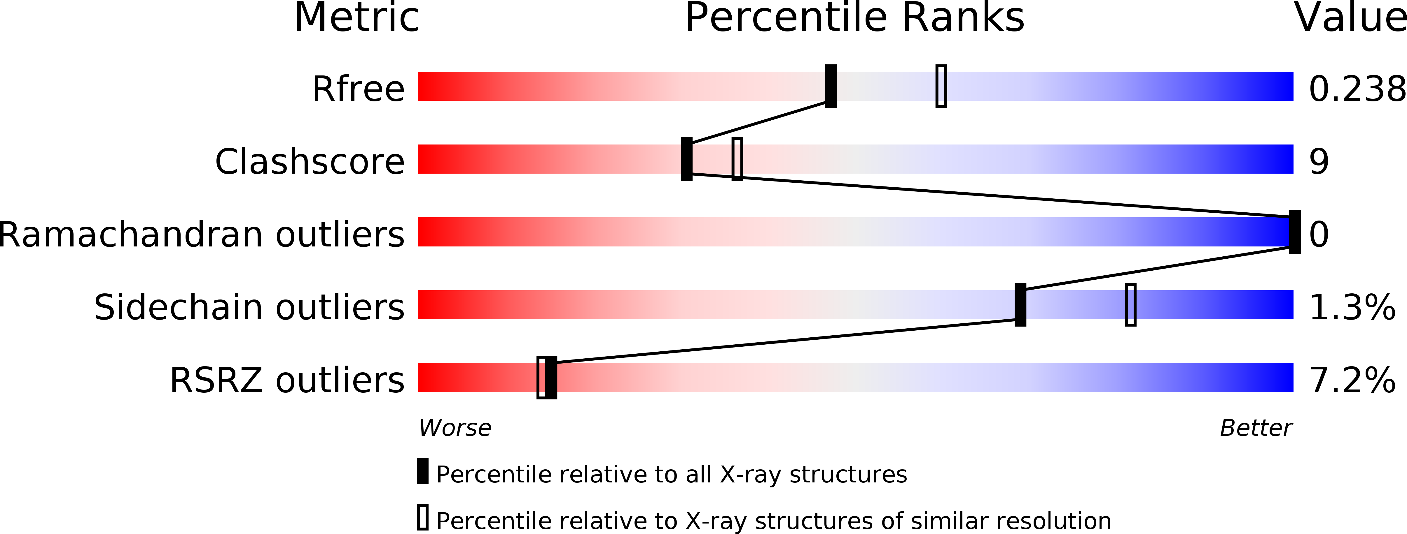
3PWE image
Deposition Date 2010-12-08
Release Date 2011-10-19
Last Version Date 2023-09-13
Entry Detail
PDB ID:
3PWE
Keywords:
Title:
Crystal structure of the E. coli beta clamp mutant R103C, I305C, C260S, C333S at 2.2A resolution
Biological Source:
Source Organism:
Host Organism:
Method Details:
Experimental Method:
Resolution:
2.20 Å
R-Value Free:
0.24
R-Value Work:
0.20
R-Value Observed:
0.20
Space Group:
P 1 21 1
Macromolecular Entities
Polymer Type:polypeptide(L)
Molecule:DNA polymerase III subunit beta
Gene (Uniprot):dnaN
Mutations:R103C, I305C, C260S, C333S
Chain IDs:A, B
Chain Length:366
Number of Molecules:2
Biological Source:Escherichia coli
Primary Citation
The E. coli clamp loader can actively pry open the beta-sliding clamp
J.Biol.Chem. 286 42704 42714 (2011)
PMID: 21971175 DOI: 10.1074/jbc.M111.268169

Abstact

Clamp loaders load ring-shaped sliding clamps onto DNA. Once loaded onto DNA, sliding clamps bind to DNA polymerases to increase the processivity of DNA synthesis. To load clamps onto DNA, an open clamp loader-clamp complex must form. An unresolved question is whether clamp loaders capture clamps that have transiently opened or whether clamp loaders bind closed clamps and actively open clamps. A simple fluorescence-based clamp opening assay was developed to address this question and to determine how ATP binding contributes to clamp opening. A direct comparison of real time binding and opening reactions revealed that the Escherichia coli γ complex binds β first and then opens the clamp. Mutation of conserved "arginine fingers" in the γ complex that interact with bound ATP decreased clamp opening activity showing that arginine fingers make an important contribution to the ATP-induced conformational changes that allow the clamp loader to pry open the clamp.

Legend

Protein

Chemical

Disease

Primary Citation of related structures