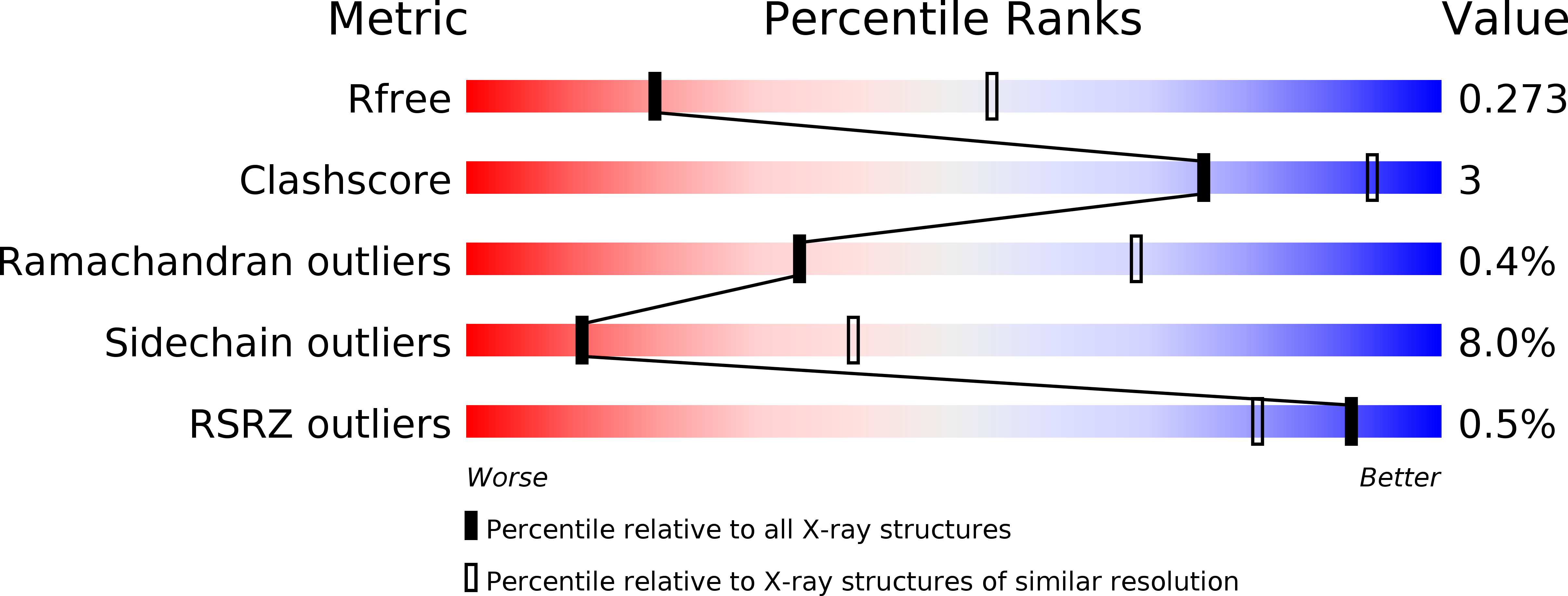
3PUF image
Deposition Date 2010-12-04
Release Date 2010-12-22
Last Version Date 2023-11-01
Entry Detail
PDB ID:
3PUF
Keywords:
Title:
Crystal structure of human RNase H2 complex
Biological Source:
Source Organism:
Homo sapiens (Taxon ID: 9606)
Host Organism:
Method Details:
Experimental Method:
Resolution:
3.10 Å
R-Value Free:
0.25
R-Value Work:
0.20
R-Value Observed:
0.20
Space Group:
P 1
Macromolecular Entities
Structures with similar UniProt ID
Protein Blast
Polymer Type:polypeptide(L)
Molecule:Ribonuclease H2 subunit A
Gene (Uniprot):RNASEH2A
Chain IDs:A, D, G, J, M, P
Chain Length:302
Number of Molecules:6
Biological Source:Homo sapiens
Structures with similar UniProt ID
Protein Blast
Polymer Type:polypeptide(L)
Molecule:Ribonuclease H2 subunit B
Gene (Uniprot):RNASEH2B
Chain IDs:B, E, H, K, N, Q
Chain Length:224
Number of Molecules:6
Biological Source:Homo sapiens
Structures with similar UniProt ID
Protein Blast
Polymer Type:polypeptide(L)
Molecule:Ribonuclease H2 subunit C
Gene (Uniprot):RNASEH2C
Chain IDs:C, F, I, L, O, R
Chain Length:167
Number of Molecules:6
Biological Source:Homo sapiens
Primary Citation
The structural and biochemical characterization of human RNase H2 complex reveals the molecular basis for substrate recognition and Aicardi-Goutieres syndrome defects.
J.Biol.Chem. 286 10540 10550 (2011)
PMID: 21177858 DOI: 10.1074/jbc.M110.181974

Abstact

RNase H2 cleaves RNA sequences that are part of RNA/DNA hybrids or that are incorporated into DNA, thus, preventing genomic instability and the accumulation of aberrant nucleic acid, which in humans induces Aicardi-Goutières syndrome, a severe autoimmune disorder. The 3.1 Å crystal structure of human RNase H2 presented here allowed us to map the positions of all 29 mutations found in Aicardi-Goutières syndrome patients, several of which were not visible in the previously reported mouse RNase H2. We propose the possible effects of these mutations on the protein stability and function. Bacterial and eukaryotic RNases H2 differ in composition and substrate specificity. Bacterial RNases H2 are monomeric proteins and homologs of the eukaryotic RNases H2 catalytic subunit, which in addition possesses two accessory proteins. The eukaryotic RNase H2 heterotrimeric complex recognizes RNA/DNA hybrids and (5')RNA-DNA(3')/DNA junction hybrids as substrates with similar efficiency, whereas bacterial RNases H2 are highly specialized in the recognition of the (5')RNA-DNA(3') junction and very poorly cleave RNA/DNA hybrids in the presence of Mg(2+) ions. Using the crystal structure of the Thermotoga maritima RNase H2-substrate complex, we modeled the human RNase H2-substrate complex and verified the model by mutational analysis. Our model indicates that the difference in substrate preference stems from the different position of the crucial tyrosine residue involved in substrate binding and recognition.

Legend

Protein

Chemical

Disease

Primary Citation of related structures