3PT2 image
Deposition Date 2010-12-02
Release Date 2011-01-19
Last Version Date 2023-05-31
Entry Detail
PDB ID:
3PT2
Title:
Structure of a viral OTU domain protease bound to Ubiquitin
Biological Source:
Source Organism:
Host Organism:
Method Details:
Experimental Method:
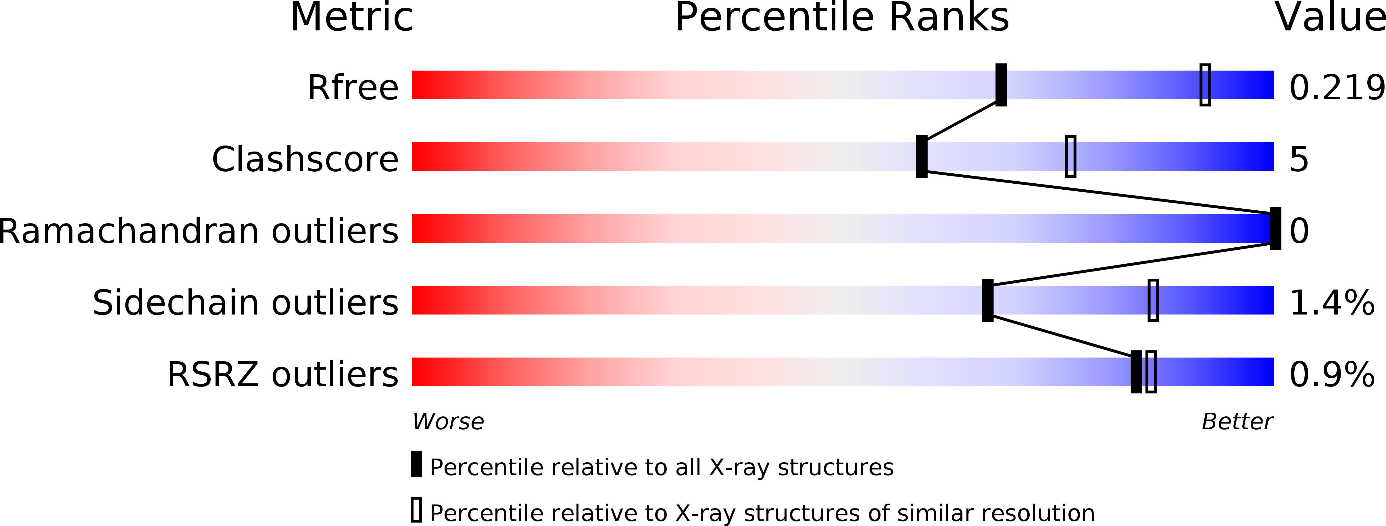
Resolution:
2.50 Å
R-Value Free:
0.22
R-Value Work:
0.16
R-Value Observed:
0.16
Space Group:
P 62 2 2
Macromolecular Entities
Polymer Type:polypeptide(L)
Molecule:RNA polymerase
Chain IDs:A
Chain Length:187
Number of Molecules:1
Biological Source:Crimean-Congo hemorrhagic fever virus
Polymer Type:polypeptide(L)
Molecule:Ubiquitin B
Chain IDs:B
Chain Length:75
Number of Molecules:1
Biological Source:Homo sapiens
Modified Residue
Compound ID Chain ID Parent Comp ID Details 2D Image
MSE A MET SELENOMETHIONINE
Primary Citation
Structural basis for the removal of ubiquitin and interferon-stimulated gene 15 by a viral ovarian tumor domain-containing protease.
Proc.Natl.Acad.Sci.USA 108 2222 2227 (2011)
PMID: 21245344 DOI: 10.1073/pnas.1013388108

Abstact

The attachment of ubiquitin (Ub) and the Ub-like (Ubl) molecule interferon-stimulated gene 15 (ISG15) to cellular proteins mediates important innate antiviral responses. Ovarian tumor (OTU) domain proteases from nairoviruses and arteriviruses were recently found to remove these molecules from host proteins, which inhibits Ub and ISG15-dependent antiviral pathways. This contrasts with the Ub-specific activity of known eukaryotic OTU-domain proteases. Here we describe crystal structures of a viral OTU domain from the highly pathogenic Crimean-Congo haemorrhagic fever virus (CCHFV) bound to Ub and to ISG15 at 2.5-Å and 2.3-Å resolution, respectively. The complexes provide a unique structural example of ISG15 bound to another protein and reveal the molecular mechanism of an ISG15 cross-reactive deubiquitinase. To accommodate structural differences between Ub and ISG15, the viral protease binds the β-grasp folds of Ub and C-terminal Ub-like domain of ISG15 in an orientation that is rotated nearly 75° with respect to that observed for Ub bound to a representative eukaryotic OTU domain from yeast. Distinct structural determinants necessary for binding either substrate were identified and allowed the reengineering of the viral OTU protease into enzymes with increased substrate specificity, either for Ub or for ISG15. Our findings now provide the basis to determine in vivo the relative contributions of deubiquitination and deISGylation to viral immune evasion tactics, and a structural template of a promiscuous deubiquitinase from a haemorrhagic fever virus that can be targeted for inhibition using small-molecule-based strategies.

Legend

Protein

Chemical

Disease

Primary Citation of related structures