3PQR image
Deposition Date 2010-11-26
Release Date 2011-03-09
Last Version Date 2024-10-30
Entry Detail
PDB ID:
3PQR
Title:
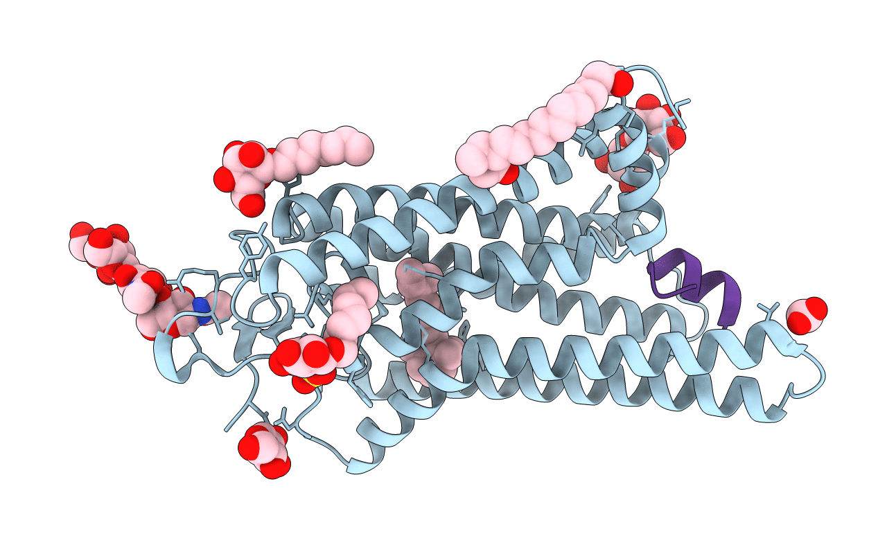
Crystal structure of Metarhodopsin II in complex with a C-terminal peptide derived from the Galpha subunit of transducin
Biological Source:
Source Organism:
Bos taurus (Taxon ID: 9913)
Method Details:
Experimental Method:
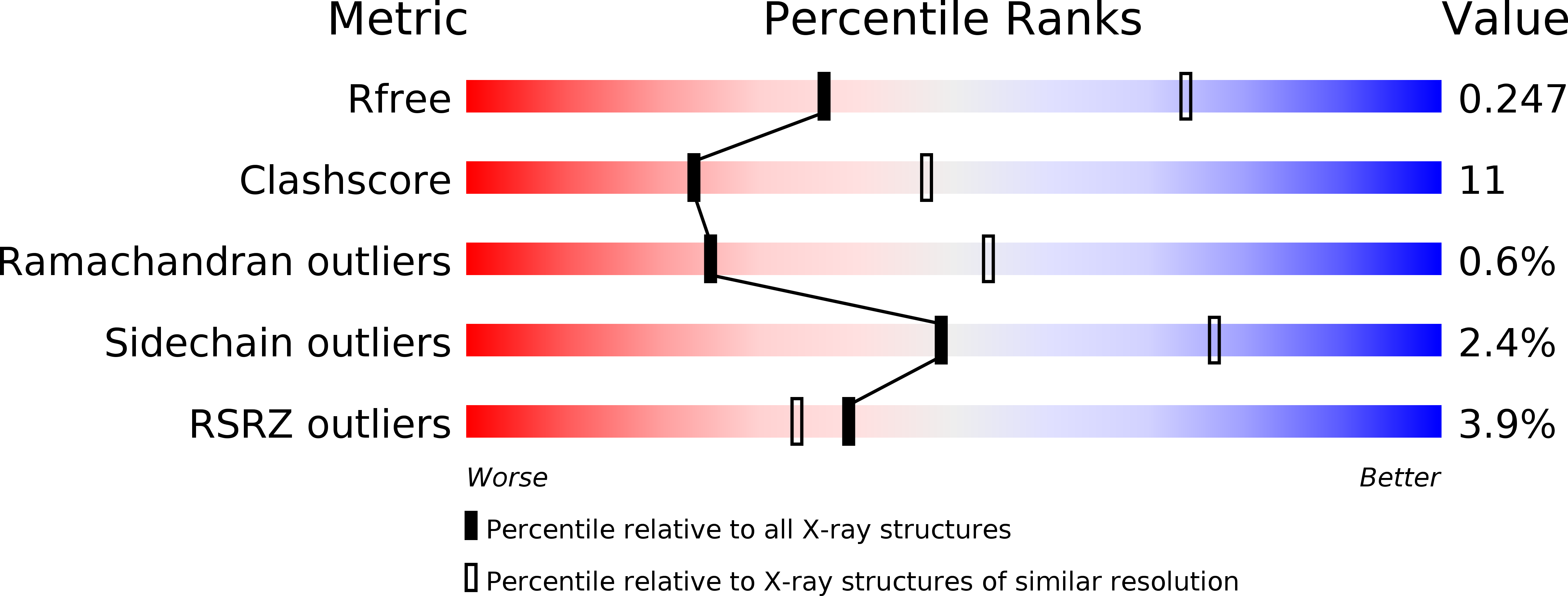
Resolution:
2.85 Å
R-Value Free:
0.24
R-Value Work:
0.21
Space Group:
H 3 2
Macromolecular Entities
Polymer Type:polypeptide(L)
Molecule:Rhodopsin
Gene (Uniprot):RHO
Chain IDs:A
Chain Length:348
Number of Molecules:1
Biological Source:Bos taurus
Polymer Type:polypeptide(L)
Molecule:Guanine nucleotide-binding protein G(t) subunit alpha-1
Gene (Uniprot):GNAT1
Mutations:K341L,C347V
Chain IDs:B
Chain Length:11
Number of Molecules:1
Biological Source:Bos taurus
Modified Residue
Compound ID Chain ID Parent Comp ID Details 2D Image
ASN A ASN GLYCOSYLATION SITE
Peptide-like Molecules
PRD_900006
Primary Citation
Crystal structure of metarhodopsin II.
Nature 471 651 655 (2011)
PMID: 21389988 DOI: 10.1038/nature09789

Abstact

G-protein-coupled receptors (GPCRs) are seven transmembrane helix (TM) proteins that transduce signals into living cells by binding extracellular ligands and coupling to intracellular heterotrimeric G proteins (Gαβγ). The photoreceptor rhodopsin couples to transducin and bears its ligand 11-cis-retinal covalently bound via a protonated Schiff base to the opsin apoprotein. Absorption of a photon causes retinal cis/trans isomerization and generates the agonist all-trans-retinal in situ. After early photoproducts, the active G-protein-binding intermediate metarhodopsin II (Meta II) is formed, in which the retinal Schiff base is still intact but deprotonated. Dissociation of the proton from the Schiff base breaks a major constraint in the protein and enables further activating steps, including an outward tilt of TM6 and formation of a large cytoplasmic crevice for uptake of the interacting C terminus of the Gα subunit. Owing to Schiff base hydrolysis, Meta II is short-lived and notoriously difficult to crystallize. We therefore soaked opsin crystals with all-trans-retinal to form Meta II, presuming that the crystal's high concentration of opsin in an active conformation (Ops*) may facilitate all-trans-retinal uptake and Schiff base formation. Here we present the 3.0 Å and 2.85 Å crystal structures, respectively, of Meta II alone or in complex with an 11-amino-acid C-terminal fragment derived from Gα (GαCT2). GαCT2 binds in a large crevice at the cytoplasmic side, akin to the binding of a similar Gα-derived peptide to Ops* (ref. 7). In the Meta II structures, the electron density from the retinal ligand seamlessly continues into the Lys 296 side chain, reflecting proper formation of the Schiff base linkage. The retinal is in a relaxed conformation and almost undistorted compared with pure crystalline all-trans-retinal. By comparison with early photoproducts we propose how retinal translocation and rotation induce the gross conformational changes characteristic for Meta II. The structures can now serve as models for the large GPCR family.

Legend

Protein

Chemical

Disease

Primary Citation of related structures