3PPG image
Deposition Date 2010-11-24
Release Date 2011-10-12
Last Version Date 2023-09-06
Entry Detail
PDB ID:
3PPG
Keywords:
Title:
Crystal structure of the Candida albicans methionine synthase by surface entropy reduction, alanine variant with zinc
Biological Source:
Source Organism:
Host Organism:
Method Details:
Experimental Method:
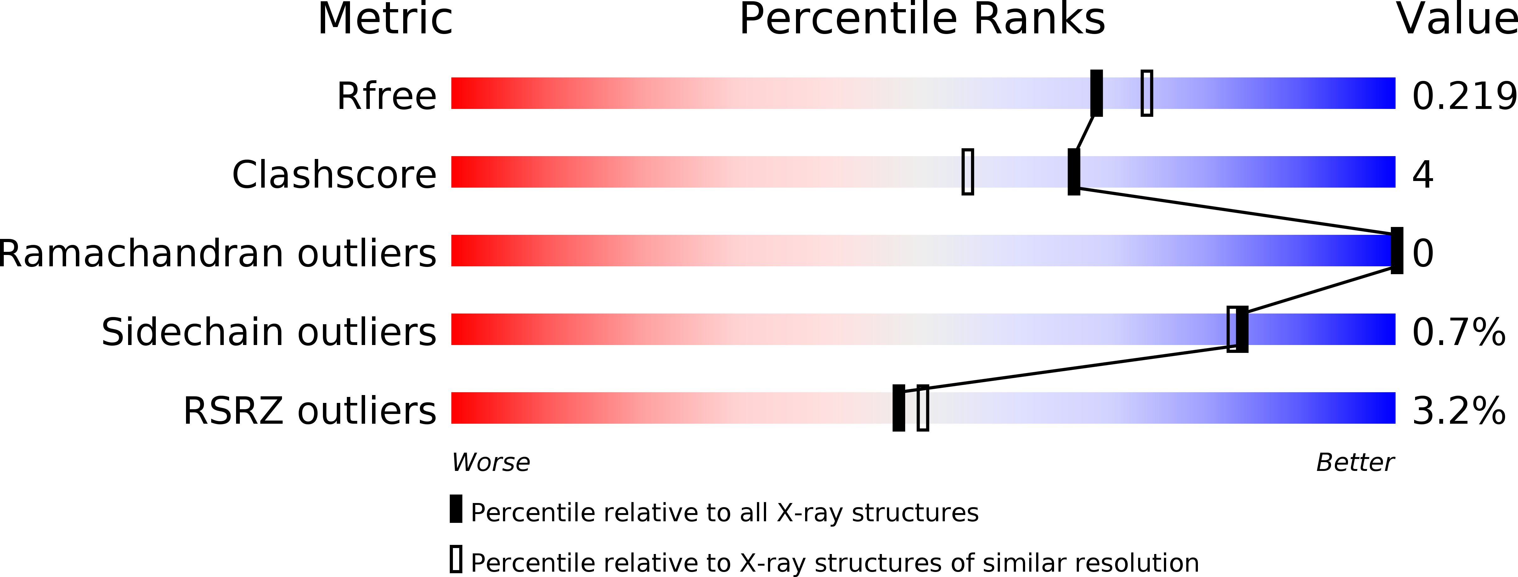
Resolution:
1.98 Å
R-Value Free:
0.22
R-Value Work:
0.17
R-Value Observed:
0.18
Space Group:
P 21 21 21
Macromolecular Entities
Polymer Type:polypeptide(L)
Molecule:5-methyltetrahydropteroyltriglutamate--homocysteine methyltransferase
Gene (Uniprot):MET6
Mutations:K103A, K104A, E107A
Chain IDs:A
Chain Length:789
Number of Molecules:1
Biological Source:Candida albicans
Ligand Molecules
Primary Citation
Structure of Candida albicans methionine synthase determined by employing surface residue mutagenesis.
Arch.Biochem.Biophys. 513 19 26 (2011)
PMID: 21689631 DOI: 10.1016/j.abb.2011.06.002

Abstact

Fungal methionine synthase, Met6p, transfers a methyl group from 5-methyl-tetrahydrofolate to homocysteine to generate methionine. The enzyme is essential to fungal growth and is a potential anti-fungal drug design target. We have characterized the enzyme from the pathogen Candida albicans but were unable to crystallize it in native form. We converted Lys103, Lys104, and Glu107 all to Tyr (Met6pY), Thr (Met6pT) and Ala (Met6pA). All variants showed wild-type kinetic activity and formed useful crystals, each with unique crystal packing. In each case the mutated residues participated in beneficial crystal contacts. We have solved the three structures at 2.0-2.8Å resolution and analyzed crystal packing, active-site residues, and similarity to other known methionine synthase structures. C. albicans Met6p has a two domain structure with each of the domains having a (βα)(8)-barrel fold. The barrels are arranged face-to-face and the active site is located in a cleft between the two domains. Met6p utilizes a zinc ion for catalysis that is bound in the C-terminal domain and ligated by four conserved residues: His657, Cys659, Glu679 and Cys739.

Legend

Protein

Chemical

Disease

Primary Citation of related structures