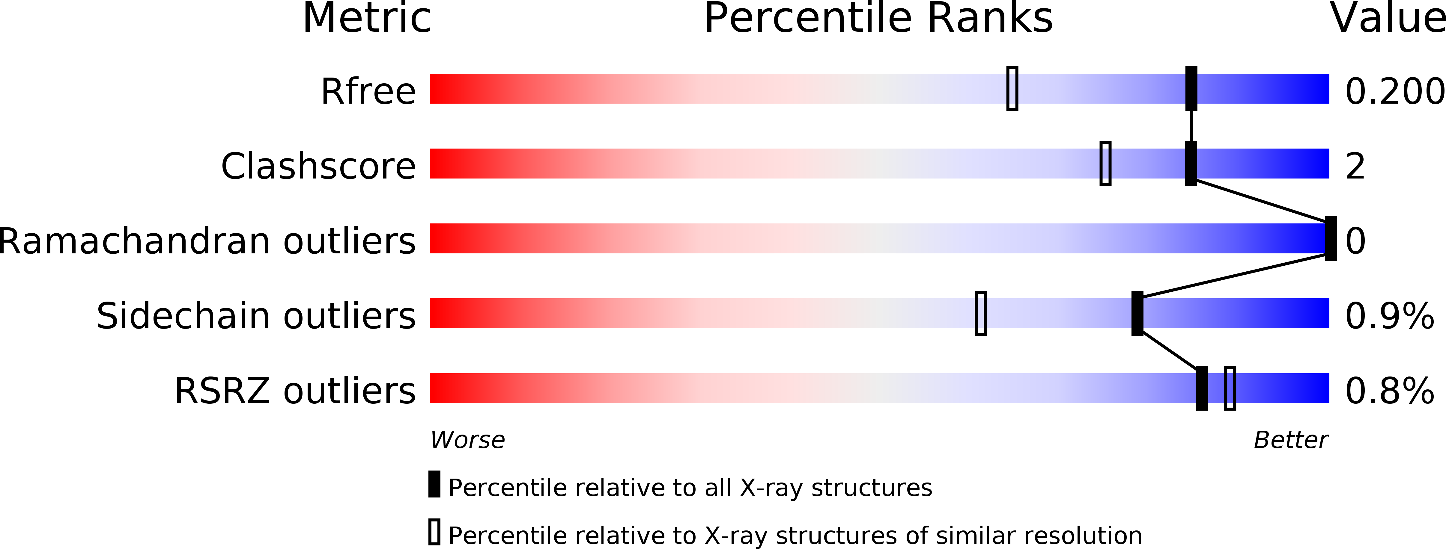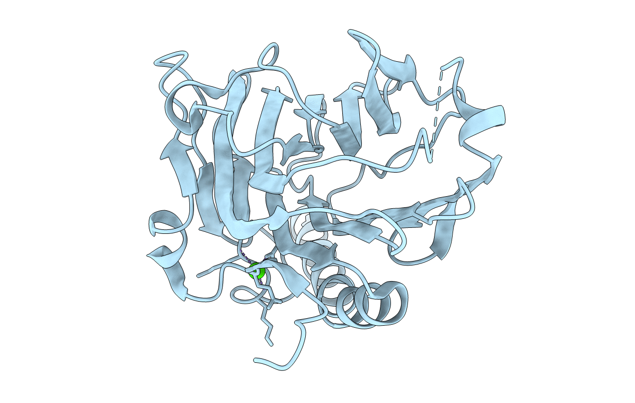
3POW image
Entry Detail
PDB ID:
3POW
Keywords:
Title:
Crystal structure of the globular domain of human calreticulin
Biological Source:
Source Organism:
Host Organism:
PDB Version:
Deposition Date:
2010-11-23
Release Date:
2011-03-09
Method Details:
Experimental Method:
Resolution:
1.55 Å
R-Value Free:
0.19
R-Value Work:
0.17
R-Value Observed:
0.17
Space Group:
P 21 21 21
Macromolecular Entities
Polymer Type:polypeptide(L)
Description:calreticulin
Chain IDs:A
Chain Length:265
Number of Molecules:1
Biological Source:Homo sapiens
Ligand Molecules
Primary Citation
X-ray structure of the human calreticulin globular domain reveals a Peptide-binding area and suggests a multi-molecular mechanism
Plos One 6 e17886 e17886 (2011)
PMID: 21423620 DOI: 10.1371/journal.pone.0017886

Abstact

In the endoplasmic reticulum, calreticulin acts as a chaperone and a Ca(2+)-signalling protein. At the cell surface, it mediates numerous important biological effects. The crystal structure of the human calreticulin globular domain was solved at 1.55 Å resolution. Interactions of the flexible N-terminal extension with the edge of the lectin site are consistently observed, revealing a hitherto unidentified peptide-binding site. A calreticulin molecular zipper, observed in all crystal lattices, could further extend this site by creating a binding cavity lined by hydrophobic residues. These data thus provide a first structural insight into the lectin-independent binding properties of calreticulin and suggest new working hypotheses, including that of a multi-molecular mechanism.

Legend

Protein

Chemical

Disease

Primary Citation of related structures