3POM image
Deposition Date 2010-11-23
Release Date 2011-04-27
Last Version Date 2024-11-06
Entry Detail
PDB ID:
3POM
Keywords:
Title:
Crystal Structure of the Unliganded Retinoblastoma Protein Pocket Domain
Biological Source:
Source Organism:
Homo sapiens (Taxon ID: 9606)
Host Organism:
Method Details:
Experimental Method:
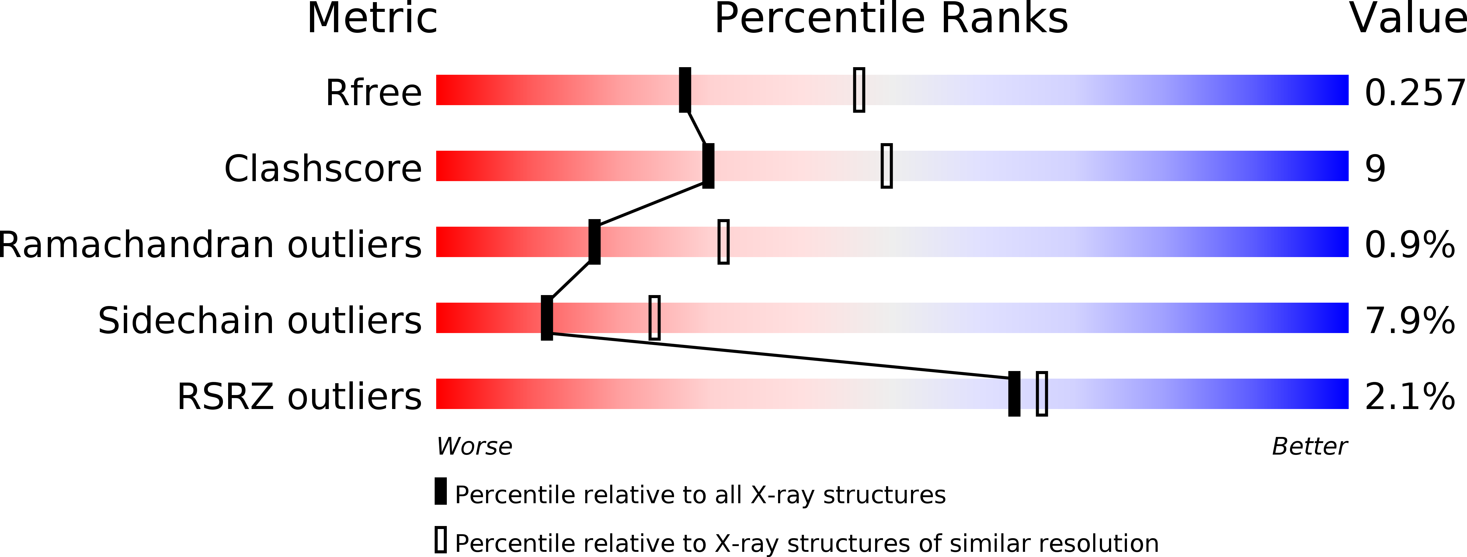
Resolution:
2.50 Å
R-Value Free:
0.26
R-Value Work:
0.21
R-Value Observed:
0.21
Space Group:
P 21 21 21
Macromolecular Entities
Structures with similar UniProt ID
Protein Blast
Polymer Type:polypeptide(L)
Molecule:Retinoblastoma-associated protein
Gene (Uniprot):RB1
Chain IDs:A, B
Chain Length:352
Number of Molecules:2
Biological Source:Homo sapiens
Primary Citation
Crystal structure of the unliganded retinoblastoma protein pocket domain.
Proteins 79 2010 2014 (2011)
PMID: 21491492 DOI: 10.1002/prot.23007

Abstact

The retinoblastoma protein (Rb) regulates cell proliferation through its association with E2F transcription factors and other proteins. The Rb “pocket” domain primarily facilitates protein-protein interactions, and several structures of the pocket bound to E2F and tumorigenic viral proteins have been reported. We report here the first crystal structure of the pocket domain without bound ligand. We find that ligand association results in observable structural changes at the binding sites but no significant changes to the overall conformation of the domain. These data support models for regulation of Rb-E2F binding that do not require considerable structural changes in the pocket domain.

Legend

Protein

Chemical

Disease

Primary Citation of related structures