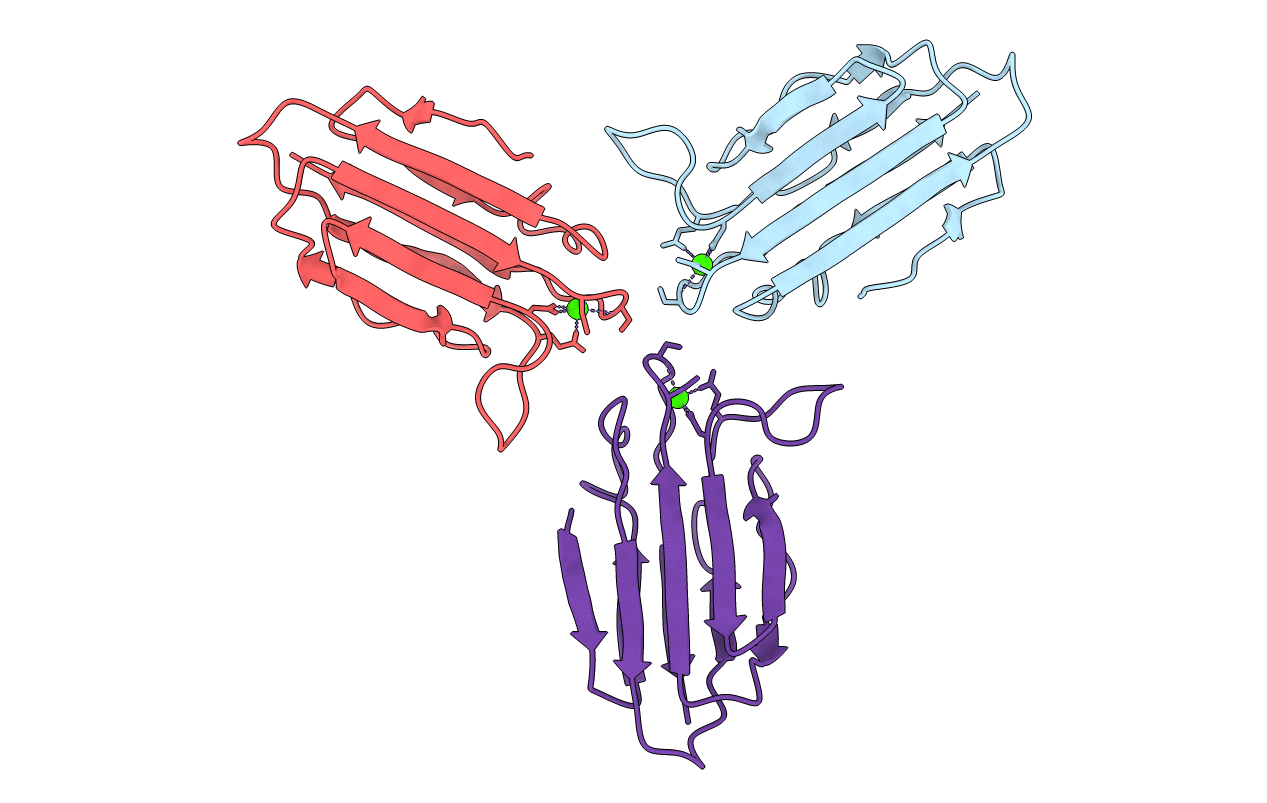
3POG image
Deposition Date 2010-11-22
Release Date 2011-08-24
Last Version Date 2024-11-20
Entry Detail
PDB ID:
3POG
Keywords:
Title:
Crystal structure of the MASP-1 CUB2 domain bound to Ca2+
Biological Source:
Source Organism:
Host Organism:
Method Details:
Experimental Method:
Resolution:
2.75 Å
R-Value Free:
0.28
R-Value Work:
0.23
R-Value Observed:
0.23
Space Group:
C 1 2 1
Macromolecular Entities
Structures with similar UniProt ID
Protein Blast
Polymer Type:polypeptide(L)
Molecule:Mannan-binding lectin serine protease 1
Gene (Uniprot):Masp1
Chain IDs:A, B, C
Chain Length:115
Number of Molecules:3
Biological Source:Rattus norvegicus
Ligand Molecules
Primary Citation
Structural Basis of Mannan-Binding Lectin Recognition by Its Associated Serine Protease MASP-1: Implications for Complement Activation.
Structure 19 1635 1643 (2011)
PMID: 22078562 DOI: 10.1016/j.str.2011.08.014

Abstact

Complement activation contributes directly to health and disease. It neutralizes pathogens and stimulates immune processes. Defects lead to immunodeficiency and autoimmune diseases, whereas inappropriate activation causes self-damage. In the lectin and classical pathways, complement is triggered upon recognition of a pathogen by an activating complex. Here we present the first structure of such a complex in the form of the collagen-like domain of mannan-binding lectin (MBL) and the binding domain of its associated protease (MASP-1/-3). The collagen binds within a groove using a pivotal lysine side chain that interacts with Ca(2+)-coordinating residues, revealing the essential role of Ca(2+). This mode of binding is prototypic for all activating complexes of the lectin and classical pathways, and suggests a general mechanism for the global changes that drive activation. The structural insights reveal a new focus for inhibitors and we have validated this concept by targeting the binding pocket of the MASP.

Legend

Protein

Chemical

Disease

Primary Citation of related structures
Feedback Form
Name
Email
Institute
Feedback